A preliminary study on diffusion-weighted imaging with background suppression and maximum intensity projection for screening internal mammary lymph node metastasis of breast cancer.
IF 1 Q4 RADIOLOGY, NUCLEAR MEDICINE & MEDICAL IMAGING
{"title":"A preliminary study on diffusion-weighted imaging with background suppression and maximum intensity projection for screening internal mammary lymph node metastasis of breast cancer.","authors":"Taiyo L Harada, Takayoshi Uematsu, Kazuaki Nakashima, Takashi Sugino, Seiichirou Nishimura, Tomomi Hayashi, Yukiko Tadokoro","doi":"10.1177/20584601251339017","DOIUrl":null,"url":null,"abstract":"<p><strong>Background: </strong>Internal mammary lymph node (IM) metastasis significantly affects prognosis. However, reports using imaging techniques have been limited. Diffusion-weighted imaging with background suppression and maximum intensity projection (DWIBS-MIP) is a sophisticated imaging method that emphasizes key signal areas such as tumors or lymph nodes in three-dimensional, providing a quick and comprehensive view immediately. Therefore, DWIBS-MIP can become a screening method for IM metastasis.</p><p><strong>Purpose: </strong>To investigates the usefulness of DWIBS-MIP for screening IM metastasis in patients undergoing preoperative breast MRI.</p><p><strong>Material and methods: </strong>Breast cancer patients with suspected IM metastasis from January 2017 to June 2024 were assessed. We evaluated the visibility and the size of IM metastasis, the long axis, short axis, and short-to-long axis (S/L) ratio of the biopsied lymph nodes at DWIBS-MIP. Patient's age, maximum diameter and hormonal status of primary cancer, breast cancer stage, the presence of axillary lymph node (Ax) metastasis, the location of the primary lesion were evaluated.</p><p><strong>Results: </strong>This study included 31 patients with IM metastasis and eight without IM metastasis. DWIBS-MIP exhibited a predictive value of 79.5% for screening IM metastasis. Between two groups, morphological characteristics of IM lymph nodes, including the long axis, short axis, and S/L ratio, demonstrated no significant differences. A higher proportion of Ax metastasis was possible in cases with IM metastasis. Other clinical data did not indicate significant differences.</p><p><strong>Conclusions: </strong>DWIBS-MIP provides an easy and effective assessment method for screening IM metastasis. The study indicates incorporating DWIBS-MIP into routine clinical practice can improve IM detection accuracy.</p>","PeriodicalId":72063,"journal":{"name":"Acta radiologica open","volume":"14 6","pages":"20584601251339017"},"PeriodicalIF":1.0000,"publicationDate":"2025-06-12","publicationTypes":"Journal Article","fieldsOfStudy":null,"isOpenAccess":false,"openAccessPdf":"https://www.ncbi.nlm.nih.gov/pmc/articles/PMC12163280/pdf/","citationCount":"0","resultStr":null,"platform":"Semanticscholar","paperid":null,"PeriodicalName":"Acta radiologica open","FirstCategoryId":"1085","ListUrlMain":"https://doi.org/10.1177/20584601251339017","RegionNum":0,"RegionCategory":null,"ArticlePicture":[],"TitleCN":null,"AbstractTextCN":null,"PMCID":null,"EPubDate":"2025/6/1 0:00:00","PubModel":"eCollection","JCR":"Q4","JCRName":"RADIOLOGY, NUCLEAR MEDICINE & MEDICAL IMAGING","Score":null,"Total":0}
引用次数: 0
Abstract
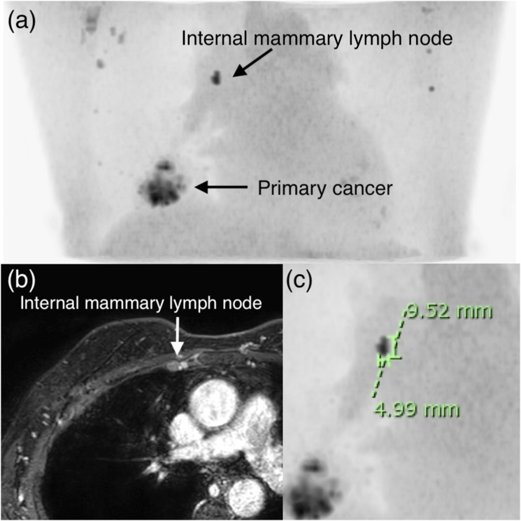
Background: Internal mammary lymph node (IM) metastasis significantly affects prognosis. However, reports using imaging techniques have been limited. Diffusion-weighted imaging with background suppression and maximum intensity projection (DWIBS-MIP) is a sophisticated imaging method that emphasizes key signal areas such as tumors or lymph nodes in three-dimensional, providing a quick and comprehensive view immediately. Therefore, DWIBS-MIP can become a screening method for IM metastasis.
Purpose: To investigates the usefulness of DWIBS-MIP for screening IM metastasis in patients undergoing preoperative breast MRI.
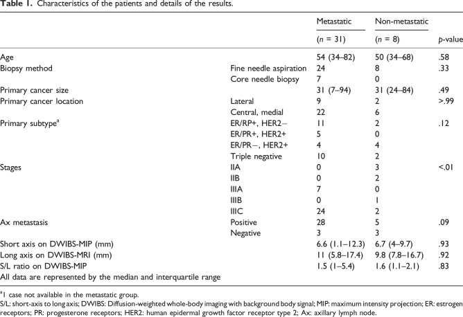
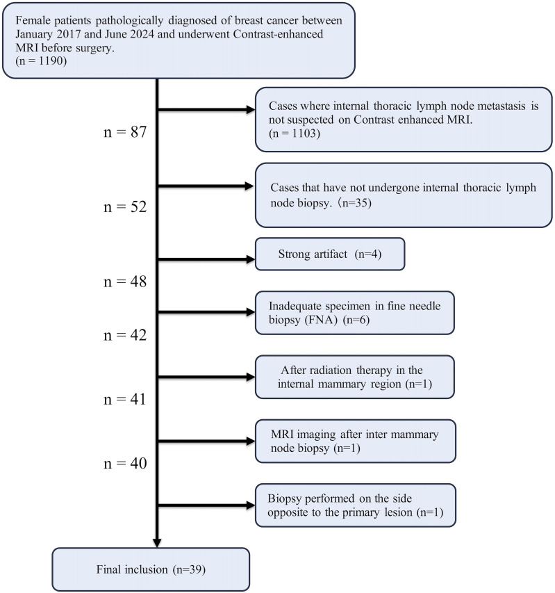
Material and methods: Breast cancer patients with suspected IM metastasis from January 2017 to June 2024 were assessed. We evaluated the visibility and the size of IM metastasis, the long axis, short axis, and short-to-long axis (S/L) ratio of the biopsied lymph nodes at DWIBS-MIP. Patient's age, maximum diameter and hormonal status of primary cancer, breast cancer stage, the presence of axillary lymph node (Ax) metastasis, the location of the primary lesion were evaluated.
Results: This study included 31 patients with IM metastasis and eight without IM metastasis. DWIBS-MIP exhibited a predictive value of 79.5% for screening IM metastasis. Between two groups, morphological characteristics of IM lymph nodes, including the long axis, short axis, and S/L ratio, demonstrated no significant differences. A higher proportion of Ax metastasis was possible in cases with IM metastasis. Other clinical data did not indicate significant differences.
Conclusions: DWIBS-MIP provides an easy and effective assessment method for screening IM metastasis. The study indicates incorporating DWIBS-MIP into routine clinical practice can improve IM detection accuracy.
背景:乳腺内淋巴结(IM)转移显著影响预后。然而,使用成像技术的报道有限。背景抑制和最大强度投影扩散加权成像(diffusion weighted imaging with background suppression and maximum intensity projection, DWIBS-MIP)是一种复杂的成像方法,可以在三维上强调肿瘤或淋巴结等关键信号区域,立即提供快速和全面的视图。因此,DWIBS-MIP可以成为一种筛查IM转移的方法。目的:探讨DWIBS-MIP在术前行乳腺MRI的患者中筛查乳腺癌转移的价值。材料与方法:对2017年1月至2024年6月疑似IM转移的乳腺癌患者进行评估。我们在DWIBS-MIP上评估了IM转移的可见性和大小,以及活检淋巴结的长轴、短轴和短长轴(S/L)比。评估患者的年龄、原发肿瘤的最大直径和激素状况、乳腺癌分期、是否存在腋窝淋巴结(Ax)转移、原发病变的位置。结果:本研究纳入了31例IM转移患者和8例未转移患者。DWIBS-MIP筛查IM转移的预测值为79.5%。两组间IM淋巴结的长轴、短轴、S/L比值等形态学特征均无显著差异。在IM转移的病例中,Ax转移的比例可能更高。其他临床数据显示无显著差异。结论:DWIBS-MIP是一种简便、有效的肿瘤转移筛查方法。研究表明,将DWIBS-MIP纳入常规临床实践可以提高IM检测的准确性。

 求助内容:
求助内容: 应助结果提醒方式:


