{"title":"Effects of platelet-rich fibrin on repair and healing chronic refractory wounds in rats by regulating Wnt/β-catenin signalling pathway.","authors":"Huasen Huang, Muyi Huang, Xinyuan Wang, Guibin Yin","doi":"10.5114/ada.2025.148762","DOIUrl":null,"url":null,"abstract":"<p><strong>Introduction: </strong>Platelet-rich fibrin is an autologous biomaterial that is rich in platelets and various growth factors, has good biocompatibility, and has significant application potential in tissue repair and regeneration.</p><p><strong>Aim: </strong>To explore the effect of platelet-rich fibrin (PRF) on the repair and healing of chronic refractory wounds in rats by regulating the Wnt/β-catenin (β-catenin) signalling pathway.</p><p><strong>Material and methods: </strong>Full-thickness skin defect open wounds were established on the backs of 50 male SD rats.</p><p><strong>Results: </strong>In comparison with the normal control group, the wound healing rate, CD34-positive cell ratio, and Wnt1, β-catenin, and c-myc protein expression levels of rats in the model group were significantly reduced at each time point; serum TNF-α, IL-1β, IL-6 levels, and GSK-3β protein expression levels were significantly increased. The granulation tissue was severely damaged and infiltrated by a large number of inflammatory cells. The collagen fibres were loosely arranged and unevenly distributed. In comparison with the model group, the wound healing rate, CD34-positive cell ratio, and Wnt1, β-catenin, and c-myc protein expression levels of rats in the PRF group and positive control group at each time point were significantly increased.</p><p><strong>Conclusions: </strong>PRF can promote the repair and healing of chronic refractory wounds in rats by inhibiting ulcer surface inflammation and promoting collagen fibre deposition and angiogenesis. Its mechanism of action may be concerned with the activation of the Wnt/β-catenin signalling pathway.</p>","PeriodicalId":54595,"journal":{"name":"Postepy Dermatologii I Alergologii","volume":"42 2","pages":"175-182"},"PeriodicalIF":1.4000,"publicationDate":"2025-04-04","publicationTypes":"Journal Article","fieldsOfStudy":null,"isOpenAccess":false,"openAccessPdf":"https://www.ncbi.nlm.nih.gov/pmc/articles/PMC12163953/pdf/","citationCount":"0","resultStr":null,"platform":"Semanticscholar","paperid":null,"PeriodicalName":"Postepy Dermatologii I Alergologii","FirstCategoryId":"3","ListUrlMain":"https://doi.org/10.5114/ada.2025.148762","RegionNum":4,"RegionCategory":"医学","ArticlePicture":[],"TitleCN":null,"AbstractTextCN":null,"PMCID":null,"EPubDate":"2025/4/1 0:00:00","PubModel":"eCollection","JCR":"Q3","JCRName":"ALLERGY","Score":null,"Total":0}
引用次数: 0
Abstract
Introduction: Platelet-rich fibrin is an autologous biomaterial that is rich in platelets and various growth factors, has good biocompatibility, and has significant application potential in tissue repair and regeneration.
Aim: To explore the effect of platelet-rich fibrin (PRF) on the repair and healing of chronic refractory wounds in rats by regulating the Wnt/β-catenin (β-catenin) signalling pathway.
Material and methods: Full-thickness skin defect open wounds were established on the backs of 50 male SD rats.
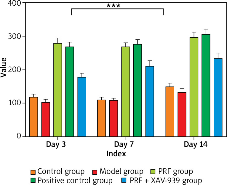
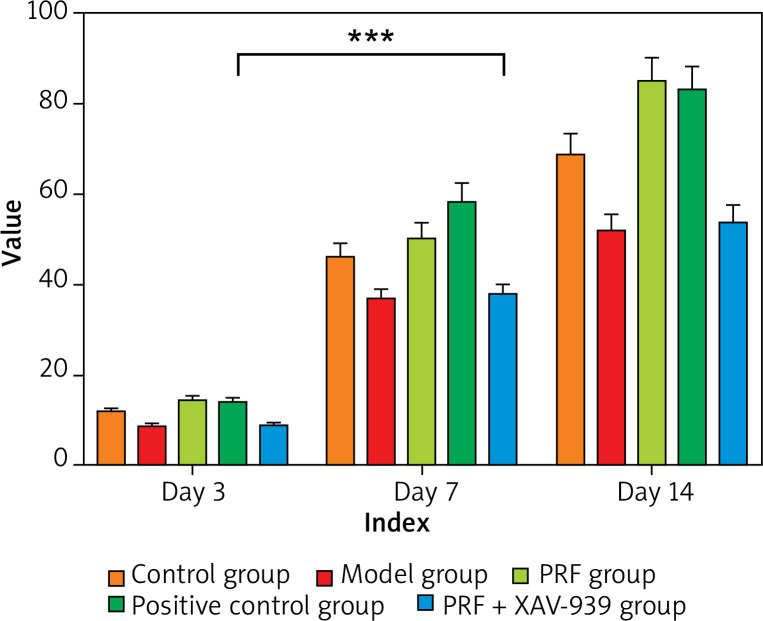
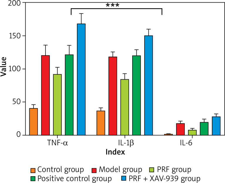
Results: In comparison with the normal control group, the wound healing rate, CD34-positive cell ratio, and Wnt1, β-catenin, and c-myc protein expression levels of rats in the model group were significantly reduced at each time point; serum TNF-α, IL-1β, IL-6 levels, and GSK-3β protein expression levels were significantly increased. The granulation tissue was severely damaged and infiltrated by a large number of inflammatory cells. The collagen fibres were loosely arranged and unevenly distributed. In comparison with the model group, the wound healing rate, CD34-positive cell ratio, and Wnt1, β-catenin, and c-myc protein expression levels of rats in the PRF group and positive control group at each time point were significantly increased.
Conclusions: PRF can promote the repair and healing of chronic refractory wounds in rats by inhibiting ulcer surface inflammation and promoting collagen fibre deposition and angiogenesis. Its mechanism of action may be concerned with the activation of the Wnt/β-catenin signalling pathway.
 求助内容:
求助内容: 应助结果提醒方式:


