Ibrahim H Yavuz, Göknur Özaydın Yavuz, Nazlı Caf, Mustafa Tümtürk, Harbiye Dilek Canat, Mehmet Onur Gökalp
{"title":"Apoptosis and PTX3 in lichen planus.","authors":"Ibrahim H Yavuz, Göknur Özaydın Yavuz, Nazlı Caf, Mustafa Tümtürk, Harbiye Dilek Canat, Mehmet Onur Gökalp","doi":"10.5114/ada.2024.145205","DOIUrl":null,"url":null,"abstract":"<p><strong>Introduction: </strong>Lichen planus (LP) is considered a T cell-mediated autoimmune disease although its aetiology remains unknown. Cytotoxic T cells play a central role in the pathogenesis of LP and these cells are known to significantly induce apoptosis in basal keratinocytes.</p><p><strong>Aim: </strong>To correlate pentraxin 3 (PTX3) with apoptosis in basal cell keratinocytes in LP and to determine whether it is a marker in this disease.</p><p><strong>Material and methods: </strong>Inclusion criteria were as follows: (i) providing written consent for participation, (ii) presence of typical clinical findings (livid-red, violaceous, flat-topped polygonal papules and plaques, fern appearance in the oral mucosa, flexural involvement, and Wickham's striae) and (iii) histopathological confirmation of LP.</p><p><strong>Results: </strong>A total of 60 participants, 30 patients and 30 controls, were included in the study. No difference was found between groups in terms of age and gender. There was a significant difference in PTX3 levels between groups. As to patients with LP, 21 (70%) patients had skin involvement only, 2 (6.7%) patients had mucosal involvement only, and 7 (23.3%) patients had both skin and mucosal involvement of LP.</p><p><strong>Conclusions: </strong>PTX3 may be associated with apoptosis in LP. Although there are no data on the usability of PTX3 as a specific marker of LP, the present study aimed to demonstrate its usability as a marker of active LP.</p>","PeriodicalId":54595,"journal":{"name":"Postepy Dermatologii I Alergologii","volume":"42 2","pages":"171-174"},"PeriodicalIF":1.4000,"publicationDate":"2024-11-21","publicationTypes":"Journal Article","fieldsOfStudy":null,"isOpenAccess":false,"openAccessPdf":"https://www.ncbi.nlm.nih.gov/pmc/articles/PMC12163958/pdf/","citationCount":"0","resultStr":null,"platform":"Semanticscholar","paperid":null,"PeriodicalName":"Postepy Dermatologii I Alergologii","FirstCategoryId":"3","ListUrlMain":"https://doi.org/10.5114/ada.2024.145205","RegionNum":4,"RegionCategory":"医学","ArticlePicture":[],"TitleCN":null,"AbstractTextCN":null,"PMCID":null,"EPubDate":"2025/4/1 0:00:00","PubModel":"eCollection","JCR":"Q3","JCRName":"ALLERGY","Score":null,"Total":0}
引用次数: 0
Abstract
Introduction: Lichen planus (LP) is considered a T cell-mediated autoimmune disease although its aetiology remains unknown. Cytotoxic T cells play a central role in the pathogenesis of LP and these cells are known to significantly induce apoptosis in basal keratinocytes.
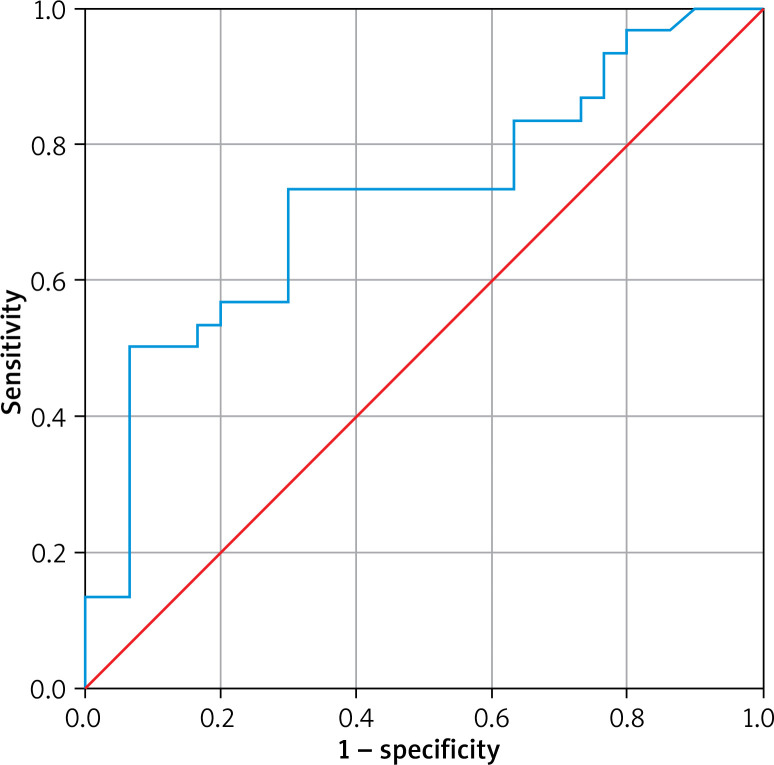
Aim: To correlate pentraxin 3 (PTX3) with apoptosis in basal cell keratinocytes in LP and to determine whether it is a marker in this disease.
Material and methods: Inclusion criteria were as follows: (i) providing written consent for participation, (ii) presence of typical clinical findings (livid-red, violaceous, flat-topped polygonal papules and plaques, fern appearance in the oral mucosa, flexural involvement, and Wickham's striae) and (iii) histopathological confirmation of LP.
Results: A total of 60 participants, 30 patients and 30 controls, were included in the study. No difference was found between groups in terms of age and gender. There was a significant difference in PTX3 levels between groups. As to patients with LP, 21 (70%) patients had skin involvement only, 2 (6.7%) patients had mucosal involvement only, and 7 (23.3%) patients had both skin and mucosal involvement of LP.
Conclusions: PTX3 may be associated with apoptosis in LP. Although there are no data on the usability of PTX3 as a specific marker of LP, the present study aimed to demonstrate its usability as a marker of active LP.
 求助内容:
求助内容: 应助结果提醒方式:


