{"title":"Morphological Markers of Chromosomal Instability: A Useful Aid in Effusion Cytology.","authors":"Archana Buch, Mallika Agarwal, Mangesh Londhe, Madhuri Singh, Akshi Raj, Charusheela Gore","doi":"10.4103/joc.joc_15_24","DOIUrl":null,"url":null,"abstract":"<p><strong>Background: </strong>Chromosomal instability (CI) is essential for carcinogenesis. Micronuclei (MN), nuclear budding (NB), chromatin bridge (CB), and multipolar mitosis (MPM) are the morphological markers of CI. These markers can be studied in the effusion fluids like pleural and ascitic fluid. This study aimed to analyze their significance in differentiating between benign and malignant pleural and ascitic effusion fluids.</p><p><strong>Materials and methods: </strong>A prospective observational study was conducted on 100 pleural and ascitic effusion fluids, out of which 71 were benign and 29 were malignant. A total of 20 malignant cases were confirmed by cell block preparation and immunohistochemistry. Leishman-stained slides were screened under oil immersion (1000×) for MN, CB, NB, and MPM. The number of cells with each of these markers were counted per 1000 cells. The data were analyzed by calculating the mean, standard deviation (SD), and Student's <i>t</i> test. A <i>P</i> value was also computed.</p><p><strong>Results: </strong>Mean (SD) of MN, CB, NB, and MPM in malignant effusion cytology were 9.01 (4.65), 0.8846 (1.07), 0.96 (1.24), and 0.92 (0097) per 1000 cells counted, whereas, the mean for benign were1.0 (0.90), 0.39 (0.54), 0.51 (0.65), and 0.50(0.73) per 1000 cells, respectively. The difference between benign and malignant effusion cytology and MN score came out to be statistically significant with a <i>P</i> value of <0.0001.</p><p><strong>Conclusion: </strong>The markers of CI help to differentiate between malignant and benign effusion cytology in low-resource settings.</p>","PeriodicalId":50217,"journal":{"name":"Journal of Cytology","volume":"42 2","pages":"95-100"},"PeriodicalIF":1.0000,"publicationDate":"2025-04-01","publicationTypes":"Journal Article","fieldsOfStudy":null,"isOpenAccess":false,"openAccessPdf":"https://www.ncbi.nlm.nih.gov/pmc/articles/PMC12165615/pdf/","citationCount":"0","resultStr":null,"platform":"Semanticscholar","paperid":null,"PeriodicalName":"Journal of Cytology","FirstCategoryId":"3","ListUrlMain":"https://doi.org/10.4103/joc.joc_15_24","RegionNum":4,"RegionCategory":"医学","ArticlePicture":[],"TitleCN":null,"AbstractTextCN":null,"PMCID":null,"EPubDate":"2025/3/28 0:00:00","PubModel":"Epub","JCR":"Q4","JCRName":"MEDICAL LABORATORY TECHNOLOGY","Score":null,"Total":0}
引用次数: 0
Abstract
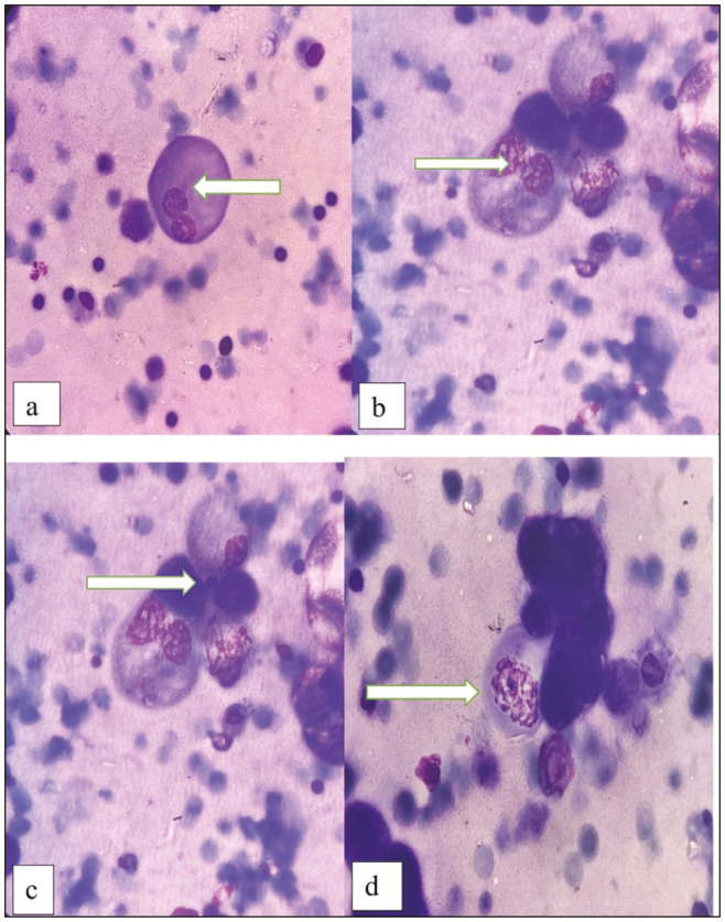
Background: Chromosomal instability (CI) is essential for carcinogenesis. Micronuclei (MN), nuclear budding (NB), chromatin bridge (CB), and multipolar mitosis (MPM) are the morphological markers of CI. These markers can be studied in the effusion fluids like pleural and ascitic fluid. This study aimed to analyze their significance in differentiating between benign and malignant pleural and ascitic effusion fluids.
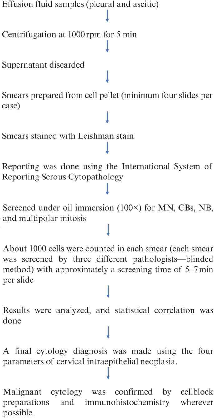
Materials and methods: A prospective observational study was conducted on 100 pleural and ascitic effusion fluids, out of which 71 were benign and 29 were malignant. A total of 20 malignant cases were confirmed by cell block preparation and immunohistochemistry. Leishman-stained slides were screened under oil immersion (1000×) for MN, CB, NB, and MPM. The number of cells with each of these markers were counted per 1000 cells. The data were analyzed by calculating the mean, standard deviation (SD), and Student's t test. A P value was also computed.
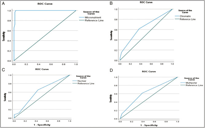
Results: Mean (SD) of MN, CB, NB, and MPM in malignant effusion cytology were 9.01 (4.65), 0.8846 (1.07), 0.96 (1.24), and 0.92 (0097) per 1000 cells counted, whereas, the mean for benign were1.0 (0.90), 0.39 (0.54), 0.51 (0.65), and 0.50(0.73) per 1000 cells, respectively. The difference between benign and malignant effusion cytology and MN score came out to be statistically significant with a P value of <0.0001.
Conclusion: The markers of CI help to differentiate between malignant and benign effusion cytology in low-resource settings.
期刊介绍:
The Journal of Cytology is the official Quarterly publication of the Indian Academy of Cytologists. It is in the 25th year of publication in the year 2008. The journal covers all aspects of diagnostic cytology, including fine needle aspiration cytology, gynecological and non-gynecological cytology. Articles on ancillary techniques, like cytochemistry, immunocytochemistry, electron microscopy, molecular cytopathology, as applied to cytological material are also welcome. The journal gives preference to clinically oriented studies over experimental and animal studies. The Journal would publish peer-reviewed original research papers, case reports, systematic reviews, meta-analysis, and debates.
 求助内容:
求助内容: 应助结果提醒方式:


