{"title":"Evaluation of Platinum Resistance or Sensitivity Markers by Analysis of Cell Block Specimens of Ascites Fluid in High-Grade Serous Carcinoma.","authors":"Rinako Ogawa, Rintaro Ohe, Takumi Kitaoka, Tsuyoshi Ohta, Satoru Nagase, Mitsuru Futakuchi","doi":"10.4103/joc.joc_152_24","DOIUrl":null,"url":null,"abstract":"<p><strong>Background: </strong>Platinum resistance in cancer cells plays an important role in the recurrence and progression of high-grade serous carcinoma (HGSC). Advanced or recurrent HGSC often presents with substantial ascites fluid retention.</p><p><strong>Aims: </strong>To demonstrate that ascites fluid sampling could help evaluate the predictive markers for platinum resistance or sensitivity in peritoneal lesions of HGSC, we compared the expression of platinum response markers of cancer cells in ascites fluid with that in peritoneal lesions.</p><p><strong>Materials and methods: </strong>Thirty samples from 10 HGSC patients were collected, including formalin-fixed paraffin-embedded specimens obtained from peritoneal lesions and primary ovarian lesions, and cell block specimens obtained from ascites fluid. The morphology of the cancer cells was evaluated by hematoxylin and eosin staining. The expression of predictive markers for platinum response in cancer cells was evaluated by immunohistochemistry for excision repair cross-complementation group 1 (ERCC1) and Schlafen 11 (SLFN11).</p><p><strong>Results: </strong>The morphology of cancer cells in cell blocks (CBs) was similar to that in peritoneal and primary lesions. The expression of ERCC1 and SLFN11 in cancer cells in CBs was positively correlated with that in peritoneal lesions. These results indicated that CBs of ascites fluid would be useful for evaluating the expression of predictive markers for platinum resistance and sensitivity.</p><p><strong>Conclusion: </strong>This study demonstrated that HGSC cells in ascites fluid may reflect the expression of predictive markers for platinum resistance or sensitivity in cancer cells in peritoneal metastatic lesions.</p>","PeriodicalId":50217,"journal":{"name":"Journal of Cytology","volume":"42 2","pages":"61-66"},"PeriodicalIF":1.0000,"publicationDate":"2025-04-01","publicationTypes":"Journal Article","fieldsOfStudy":null,"isOpenAccess":false,"openAccessPdf":"https://www.ncbi.nlm.nih.gov/pmc/articles/PMC12165613/pdf/","citationCount":"0","resultStr":null,"platform":"Semanticscholar","paperid":null,"PeriodicalName":"Journal of Cytology","FirstCategoryId":"3","ListUrlMain":"https://doi.org/10.4103/joc.joc_152_24","RegionNum":4,"RegionCategory":"医学","ArticlePicture":[],"TitleCN":null,"AbstractTextCN":null,"PMCID":null,"EPubDate":"2025/5/29 0:00:00","PubModel":"Epub","JCR":"Q4","JCRName":"MEDICAL LABORATORY TECHNOLOGY","Score":null,"Total":0}
引用次数: 0
Abstract
Background: Platinum resistance in cancer cells plays an important role in the recurrence and progression of high-grade serous carcinoma (HGSC). Advanced or recurrent HGSC often presents with substantial ascites fluid retention.
Aims: To demonstrate that ascites fluid sampling could help evaluate the predictive markers for platinum resistance or sensitivity in peritoneal lesions of HGSC, we compared the expression of platinum response markers of cancer cells in ascites fluid with that in peritoneal lesions.
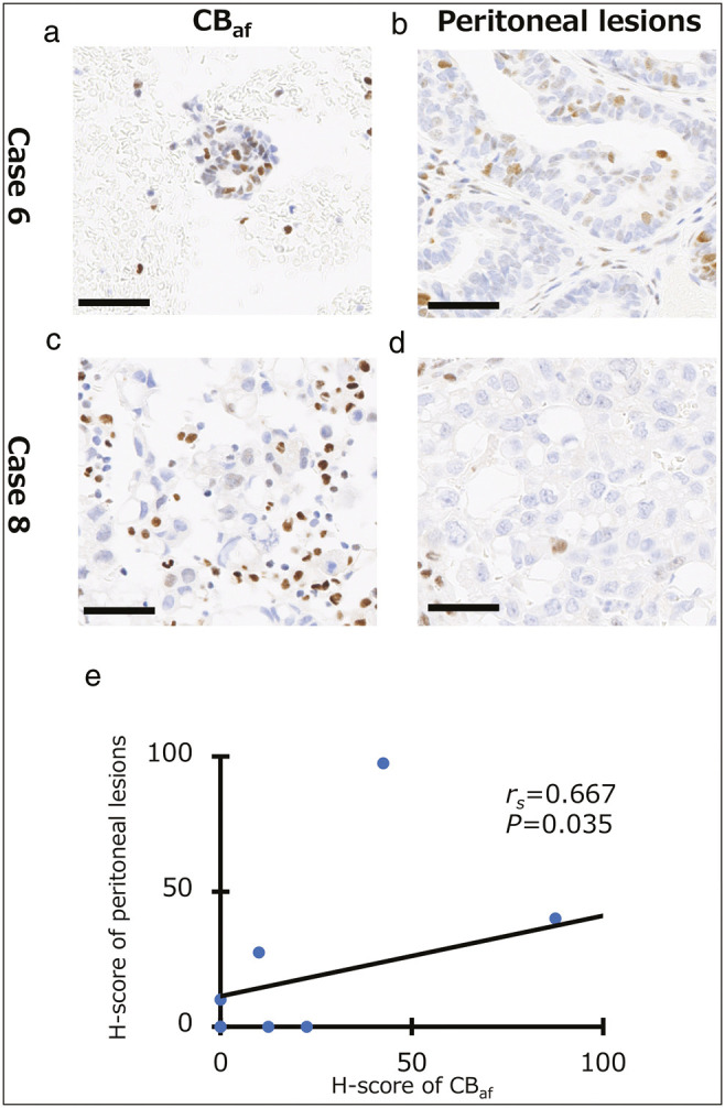
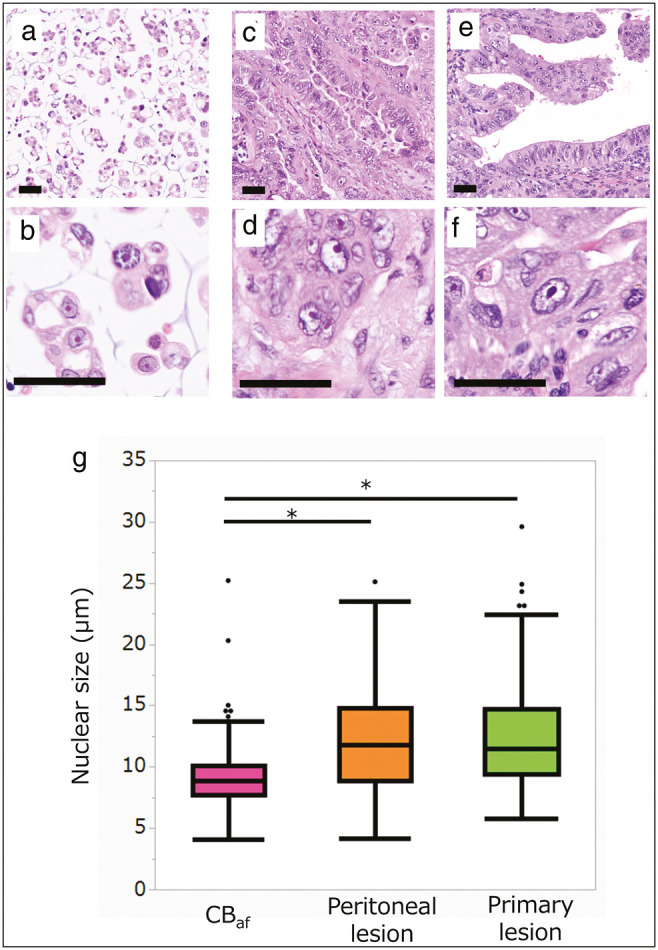
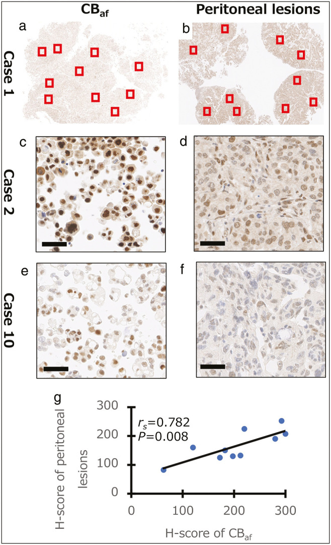
Materials and methods: Thirty samples from 10 HGSC patients were collected, including formalin-fixed paraffin-embedded specimens obtained from peritoneal lesions and primary ovarian lesions, and cell block specimens obtained from ascites fluid. The morphology of the cancer cells was evaluated by hematoxylin and eosin staining. The expression of predictive markers for platinum response in cancer cells was evaluated by immunohistochemistry for excision repair cross-complementation group 1 (ERCC1) and Schlafen 11 (SLFN11).
Results: The morphology of cancer cells in cell blocks (CBs) was similar to that in peritoneal and primary lesions. The expression of ERCC1 and SLFN11 in cancer cells in CBs was positively correlated with that in peritoneal lesions. These results indicated that CBs of ascites fluid would be useful for evaluating the expression of predictive markers for platinum resistance and sensitivity.
Conclusion: This study demonstrated that HGSC cells in ascites fluid may reflect the expression of predictive markers for platinum resistance or sensitivity in cancer cells in peritoneal metastatic lesions.
期刊介绍:
The Journal of Cytology is the official Quarterly publication of the Indian Academy of Cytologists. It is in the 25th year of publication in the year 2008. The journal covers all aspects of diagnostic cytology, including fine needle aspiration cytology, gynecological and non-gynecological cytology. Articles on ancillary techniques, like cytochemistry, immunocytochemistry, electron microscopy, molecular cytopathology, as applied to cytological material are also welcome. The journal gives preference to clinically oriented studies over experimental and animal studies. The Journal would publish peer-reviewed original research papers, case reports, systematic reviews, meta-analysis, and debates.
 求助内容:
求助内容: 应助结果提醒方式:


