Moschoula Passali, Maria Højberg Knudsen, Knud Josefsen, Julie Christine Antvorskov, Amalie Monberg Hindsholm, Ulrich Lindberg, Jette Lautrup Frederiksen, Henrik Bo Wiberg Larsson, Stig Præstekjær Cramer
{"title":"Blood-brain barrier permeability in relation to disease severity and timing of multiple sclerosis diagnosis in optic neuritis.","authors":"Moschoula Passali, Maria Højberg Knudsen, Knud Josefsen, Julie Christine Antvorskov, Amalie Monberg Hindsholm, Ulrich Lindberg, Jette Lautrup Frederiksen, Henrik Bo Wiberg Larsson, Stig Præstekjær Cramer","doi":"10.1177/20552173251346979","DOIUrl":null,"url":null,"abstract":"<p><strong>Background: </strong>Dynamic contrast-enhanced magnetic resonance imaging is a promising biomarker allowing for in vivo quantification of blood-brain barrier permeability.</p><p><strong>Objectives: </strong>To explore the relationship between blood-brain barrier permeability, optic neuritis disease severity, and multiple sclerosis conversion in optic neuritis.</p><p><strong>Methods: </strong>Gjedde-Patlak models from dynamic contrast-enhanced magnetic resonance imaging were used to estimate blood-brain barrier permeability (<i>K<sub>i</sub></i> ) in 78 optic neuritis patients. The 2017 McDonald criteria were used to diagnose multiple sclerosis with a minimum follow-up time of 2 years.</p><p><strong>Results: </strong>Normal-appearing white matter <i>K<sub>i</sub></i> correlated with the number of magnetic resonance imaging criteria for dissemination in space (Spearman's <i>ρ</i> = 0.3, <i>p</i> = 0.0074), but not with visual acuity, color vision, and inter-eye difference in retinal nerve fiber layer thickness. Normal-appearing white matter <i>K<sub>i</sub></i> did not differ between patients with and without oligoclonal bands (<i>p</i> = 0.067), but patients with brain contrast-enhancing lesions had higher normal-appearing white matter <i>K<sub>i</sub></i> than those without (<i>p</i> = 0.04). Early multiple sclerosis-converters diagnosed at optic neuritis onset (<i>n</i> = 36) had higher normal-appearing white matter <i>K<sub>i</sub></i> than non-converters (<i>n</i> = 29) (<i>p</i> = 0.01), but this was not the case for late multiple sclerosis-converters (<i>n</i> = 13) (<i>p</i> = 0.57). Normal-appearing white matter <i>K<sub>i</sub></i> did not significantly predict overall multiple sclerosis conversion (<i>p</i> = 0.068, AUC = 0.652).</p><p><strong>Conclusions: </strong>Normal-appearing white matter <i>K<sub>i</sub></i> was associated with magnetic resonance imaging biomarkers of multiple sclerosis, but not with biomarkers of optic neuritis disease severity. Normal-appearing white matter <i>K<sub>i</sub></i> was increased at, but not before, the multiple sclerosis diagnosis.</p>","PeriodicalId":18961,"journal":{"name":"Multiple Sclerosis Journal - Experimental, Translational and Clinical","volume":"11 2","pages":"20552173251346979"},"PeriodicalIF":2.3000,"publicationDate":"2025-06-11","publicationTypes":"Journal Article","fieldsOfStudy":null,"isOpenAccess":false,"openAccessPdf":"https://www.ncbi.nlm.nih.gov/pmc/articles/PMC12163281/pdf/","citationCount":"0","resultStr":null,"platform":"Semanticscholar","paperid":null,"PeriodicalName":"Multiple Sclerosis Journal - Experimental, Translational and Clinical","FirstCategoryId":"1085","ListUrlMain":"https://doi.org/10.1177/20552173251346979","RegionNum":0,"RegionCategory":null,"ArticlePicture":[],"TitleCN":null,"AbstractTextCN":null,"PMCID":null,"EPubDate":"2025/4/1 0:00:00","PubModel":"eCollection","JCR":"Q2","JCRName":"CLINICAL NEUROLOGY","Score":null,"Total":0}
引用次数: 0
Abstract
Background: Dynamic contrast-enhanced magnetic resonance imaging is a promising biomarker allowing for in vivo quantification of blood-brain barrier permeability.
Objectives: To explore the relationship between blood-brain barrier permeability, optic neuritis disease severity, and multiple sclerosis conversion in optic neuritis.
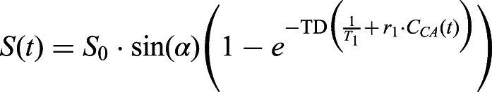
Methods: Gjedde-Patlak models from dynamic contrast-enhanced magnetic resonance imaging were used to estimate blood-brain barrier permeability (Ki ) in 78 optic neuritis patients. The 2017 McDonald criteria were used to diagnose multiple sclerosis with a minimum follow-up time of 2 years.
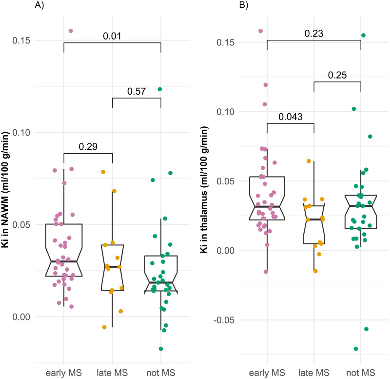
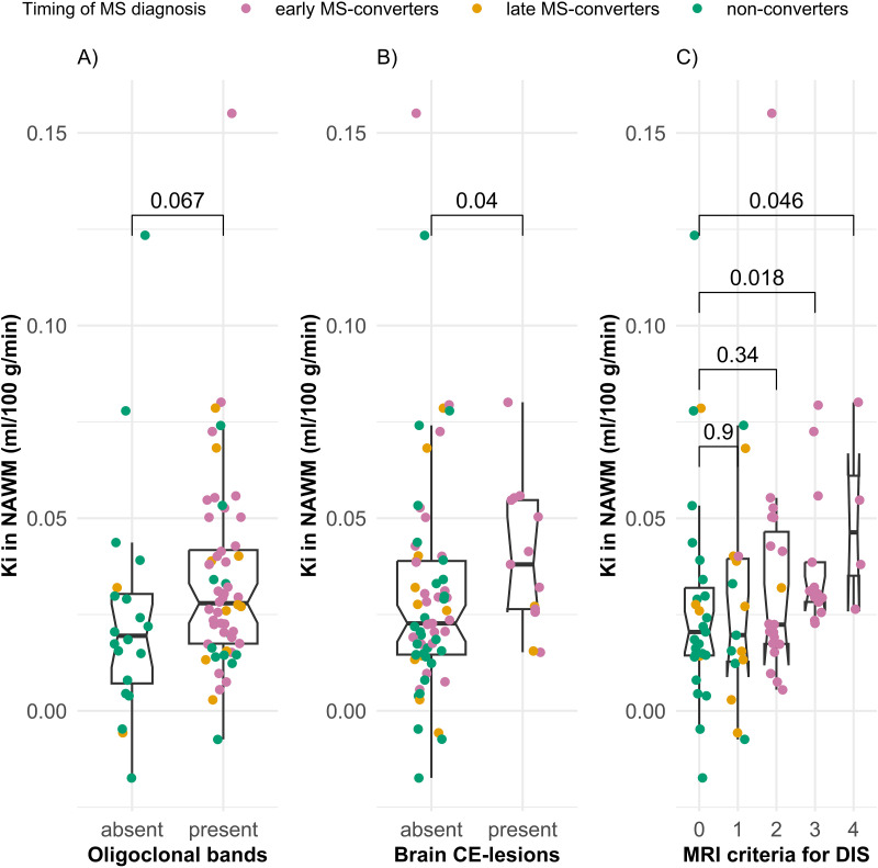
Results: Normal-appearing white matter Ki correlated with the number of magnetic resonance imaging criteria for dissemination in space (Spearman's ρ = 0.3, p = 0.0074), but not with visual acuity, color vision, and inter-eye difference in retinal nerve fiber layer thickness. Normal-appearing white matter Ki did not differ between patients with and without oligoclonal bands (p = 0.067), but patients with brain contrast-enhancing lesions had higher normal-appearing white matter Ki than those without (p = 0.04). Early multiple sclerosis-converters diagnosed at optic neuritis onset (n = 36) had higher normal-appearing white matter Ki than non-converters (n = 29) (p = 0.01), but this was not the case for late multiple sclerosis-converters (n = 13) (p = 0.57). Normal-appearing white matter Ki did not significantly predict overall multiple sclerosis conversion (p = 0.068, AUC = 0.652).
Conclusions: Normal-appearing white matter Ki was associated with magnetic resonance imaging biomarkers of multiple sclerosis, but not with biomarkers of optic neuritis disease severity. Normal-appearing white matter Ki was increased at, but not before, the multiple sclerosis diagnosis.

 求助内容:
求助内容: 应助结果提醒方式:


