David Leonardo Cruvinel Isaac, Alexandre Caiado Ferreira Pires, Laís Lauria Neves, Jamil Miguel Neto, Heitor do Amaral Simões, Karime Fugihara Iwamoto, Raphael Toledo Remiggi, Leticia Pinheiro de Freitas, Alexandre Chater Taleb, Marcos Avila
{"title":"Effect of disease duration on foveal microvasculature assessed by OCTA in type 2 diabetes mellitus without clinical diabetic retinopathy.","authors":"David Leonardo Cruvinel Isaac, Alexandre Caiado Ferreira Pires, Laís Lauria Neves, Jamil Miguel Neto, Heitor do Amaral Simões, Karime Fugihara Iwamoto, Raphael Toledo Remiggi, Leticia Pinheiro de Freitas, Alexandre Chater Taleb, Marcos Avila","doi":"10.1186/s40942-025-00694-1","DOIUrl":null,"url":null,"abstract":"<p><strong>Background: </strong>The objective of this study was to establish a comparison between the vessel density (VD) and foveal avascular zone (FAZ) of patients with type 2 diabetes mellitus (T2DM) who lacked clinical signs of diabetic retinopathy (DR) and non-diabetic patients using optical coherence tomography angiography (OCTA).</p><p><strong>Methods: </strong>A cross-sectional comparative case-control study (unpaired) was carried out at two tertiary hospitals. All subjects underwent optical coherence tomography angiography (OCTA) examination (DRI OCT Triton Swept Source, Topcon, Japan). The average VD in the superficial capillary plexus (SCP) and the deep capillary plexus (DCP), the FAZ area (mm2) in SCP, and DCP were taken into analysis. The time since the diagnosis of T2DM was used to stratify patients with diabetes between 5 and 10 years and those with a diagnosis of more than 10 years.</p><p><strong>Results: </strong>Compared to non-diabetic controls, the parafoveal VD in both SCP and DCP was significantly reduced in the eyes of T2DM patients without clinical DR (p < 0.001). Additionally, the VD was also statistically reduced in T2DM diagnosed more than 10 years ago compared to T2DM cases diagnosed between 5 and 10 years ago (p < 0.001). The FAZ area in both plexuses was larger in T2DM eyes compared to controls (p < 0.001). The FAZ area was enlarged in DCP (p = 0.04), but there was no significance of FAZ area in SCP when comparing patients with T2DM diagnosed between 5 and 10 years ago to those diagnosed more than 10 years ago (p = 0.06).</p><p><strong>Conclusion: </strong>In diabetic patients with long-term diagnosed disease, OCTA was shown to be capable of detecting preclinical microvascular foveal abnormalities prior to the development of clinically apparent retinopathy. According to our findings, OCTA has the potential to be a promising instrument for the early detection of vascular micro-abnormalities and the routine screening of diabetic eyes.</p>","PeriodicalId":14289,"journal":{"name":"International Journal of Retina and Vitreous","volume":"11 1","pages":"66"},"PeriodicalIF":2.4000,"publicationDate":"2025-06-15","publicationTypes":"Journal Article","fieldsOfStudy":null,"isOpenAccess":false,"openAccessPdf":"https://www.ncbi.nlm.nih.gov/pmc/articles/PMC12168325/pdf/","citationCount":"0","resultStr":null,"platform":"Semanticscholar","paperid":null,"PeriodicalName":"International Journal of Retina and Vitreous","FirstCategoryId":"1085","ListUrlMain":"https://doi.org/10.1186/s40942-025-00694-1","RegionNum":0,"RegionCategory":null,"ArticlePicture":[],"TitleCN":null,"AbstractTextCN":null,"PMCID":null,"EPubDate":"","PubModel":"","JCR":"Q2","JCRName":"OPHTHALMOLOGY","Score":null,"Total":0}
引用次数: 0
Abstract
Background: The objective of this study was to establish a comparison between the vessel density (VD) and foveal avascular zone (FAZ) of patients with type 2 diabetes mellitus (T2DM) who lacked clinical signs of diabetic retinopathy (DR) and non-diabetic patients using optical coherence tomography angiography (OCTA).
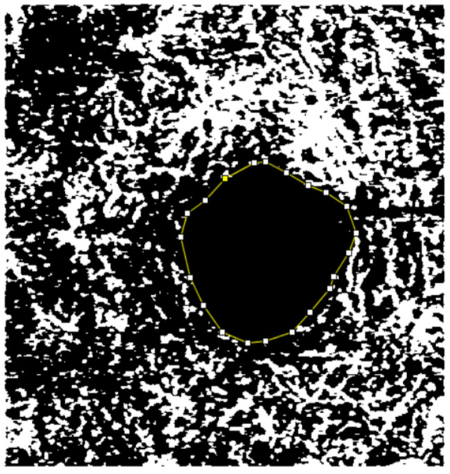
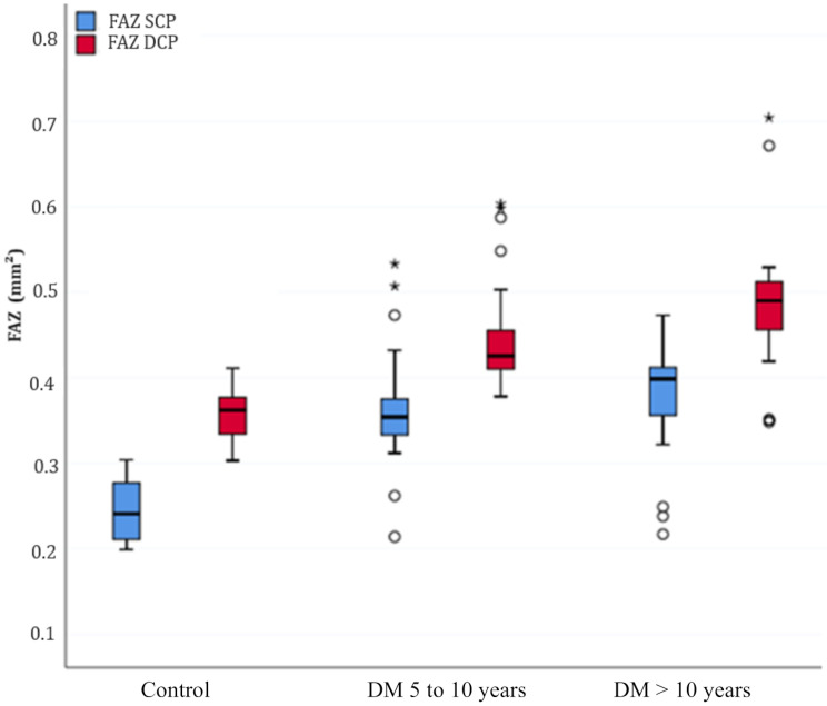
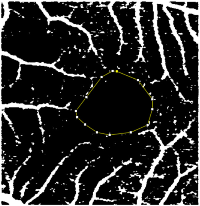
Methods: A cross-sectional comparative case-control study (unpaired) was carried out at two tertiary hospitals. All subjects underwent optical coherence tomography angiography (OCTA) examination (DRI OCT Triton Swept Source, Topcon, Japan). The average VD in the superficial capillary plexus (SCP) and the deep capillary plexus (DCP), the FAZ area (mm2) in SCP, and DCP were taken into analysis. The time since the diagnosis of T2DM was used to stratify patients with diabetes between 5 and 10 years and those with a diagnosis of more than 10 years.
Results: Compared to non-diabetic controls, the parafoveal VD in both SCP and DCP was significantly reduced in the eyes of T2DM patients without clinical DR (p < 0.001). Additionally, the VD was also statistically reduced in T2DM diagnosed more than 10 years ago compared to T2DM cases diagnosed between 5 and 10 years ago (p < 0.001). The FAZ area in both plexuses was larger in T2DM eyes compared to controls (p < 0.001). The FAZ area was enlarged in DCP (p = 0.04), but there was no significance of FAZ area in SCP when comparing patients with T2DM diagnosed between 5 and 10 years ago to those diagnosed more than 10 years ago (p = 0.06).
Conclusion: In diabetic patients with long-term diagnosed disease, OCTA was shown to be capable of detecting preclinical microvascular foveal abnormalities prior to the development of clinically apparent retinopathy. According to our findings, OCTA has the potential to be a promising instrument for the early detection of vascular micro-abnormalities and the routine screening of diabetic eyes.
背景:本研究的目的是利用光学相干断层扫描血管造影(OCTA)对无糖尿病视网膜病变(DR)临床体征的2型糖尿病(T2DM)患者和非糖尿病患者的血管密度(VD)和中央凹无血管区(FAZ)进行比较。方法:在两所三级医院进行横断面比较病例对照研究(未配对)。所有受试者接受光学相干断层扫描血管造影(OCTA)检查(DRI OCT Triton扫描源,Topcon,日本)。分析浅毛细血管丛(SCP)和深毛细血管丛(DCP)的平均VD、浅毛细血管丛FAZ面积(mm2)和深毛细血管丛。自诊断为T2DM以来的时间用于对5 - 10年的糖尿病患者和诊断超过10年的糖尿病患者进行分层。结果:与非糖尿病对照组相比,无临床DR的T2DM患者的眼中央凹旁VD在SCP和DCP中均显著降低(p结论:在长期诊断疾病的糖尿病患者中,OCTA能够在临床明显视网膜病变发生之前检测到临床前微血管中央凹异常。根据我们的研究结果,OCTA有潜力成为早期发现血管微异常和糖尿病眼常规筛查的一种有前途的工具。
期刊介绍:
International Journal of Retina and Vitreous focuses on the ophthalmic subspecialty of vitreoretinal disorders. The journal presents original articles on new approaches to diagnosis, outcomes of clinical trials, innovations in pharmacological therapy and surgical techniques, as well as basic science advances that impact clinical practice. Topical areas include, but are not limited to: -Imaging of the retina, choroid and vitreous -Innovations in optical coherence tomography (OCT) -Small-gauge vitrectomy, retinal detachment, chromovitrectomy -Electroretinography (ERG), microperimetry, other functional tests -Intraocular tumors -Retinal pharmacotherapy & drug delivery -Diabetic retinopathy & other vascular diseases -Age-related macular degeneration (AMD) & other macular entities

 求助内容:
求助内容: 应助结果提醒方式:


