{"title":"Clinical features and outcomes in pediatric patients with cortical laminar necrosis: a single-center retrospective study.","authors":"Zhanwei Zhang, Zou Pan, Lifen Yang, Fang He, Fangyun Liu, Jing Peng","doi":"10.1186/s13052-025-02038-z","DOIUrl":null,"url":null,"abstract":"<p><strong>Background: </strong>Cortical laminar necrosis (CLN) is a selective cortical necrosis primarily involving layer 3 of the cortex. Its causes, clinical features, and outcomes in pediatric patients remain unclear due to limited cases.</p><p><strong>Methods: </strong>Pediatric CLN patients from Xiangya Hospital from January 2014 to December 2024 were enrolled. Clinical data, brain magnetic resonance imaging (MRI), and Modified Rankin Scale (mRS) for neurological outcomes were analyzed.</p><p><strong>Results: </strong>Among 59 enrolled patients, causes included hypoxic-ischemic encephalopathy (HIE), hereditary and metabolic, infectious, vascular, and trauma. Seizures and consciousness changes were common symptoms. Infectious and hereditary and metabolic causes were linked to status epilepticus (P < 0.001), while vascular causes were associated with hemiplegia (P < 0.001). HIE patients had poorer prognosis (P = 0.038). MRI showed earlier hyperintensity on diffusion weighted imaging (DWI) than enhancement in T1 contrasted sequence, as well as T1 sequence and fluid attenuated inversion recovery (FLAIR). Bilateral involvement was seen in all HIE cases, while hereditary and metabolic causes often showed unilateral involvement (P < 0.001). Frontal lobe involvement was common in vascular and trauma cases (P = 0.017), and all patients with HIE exhibited involvement in the parietal and occipital lobes. While infectious cases frequently involved the insular and temporal lobes, most patients exhibited brain atrophy/encephalomalacia at follow-up. Three patients showed new CLN and two patients exhibited persistent CLN during MRI follow-up.</p><p><strong>Conclusion: </strong>HIE, hereditary/metabolic, infectious, vascular, and trauma are the main causes of pediatric CLN, with distinct clinical features and outcomes.</p>","PeriodicalId":14511,"journal":{"name":"Italian Journal of Pediatrics","volume":"51 1","pages":"188"},"PeriodicalIF":3.1000,"publicationDate":"2025-06-15","publicationTypes":"Journal Article","fieldsOfStudy":null,"isOpenAccess":false,"openAccessPdf":"https://www.ncbi.nlm.nih.gov/pmc/articles/PMC12168347/pdf/","citationCount":"0","resultStr":null,"platform":"Semanticscholar","paperid":null,"PeriodicalName":"Italian Journal of Pediatrics","FirstCategoryId":"3","ListUrlMain":"https://doi.org/10.1186/s13052-025-02038-z","RegionNum":3,"RegionCategory":"医学","ArticlePicture":[],"TitleCN":null,"AbstractTextCN":null,"PMCID":null,"EPubDate":"","PubModel":"","JCR":"Q1","JCRName":"PEDIATRICS","Score":null,"Total":0}
引用次数: 0
Abstract
Background: Cortical laminar necrosis (CLN) is a selective cortical necrosis primarily involving layer 3 of the cortex. Its causes, clinical features, and outcomes in pediatric patients remain unclear due to limited cases.
Methods: Pediatric CLN patients from Xiangya Hospital from January 2014 to December 2024 were enrolled. Clinical data, brain magnetic resonance imaging (MRI), and Modified Rankin Scale (mRS) for neurological outcomes were analyzed.
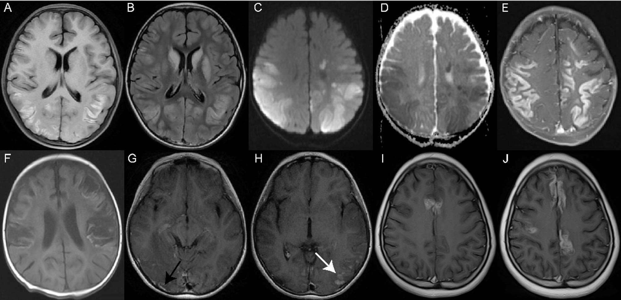
Results: Among 59 enrolled patients, causes included hypoxic-ischemic encephalopathy (HIE), hereditary and metabolic, infectious, vascular, and trauma. Seizures and consciousness changes were common symptoms. Infectious and hereditary and metabolic causes were linked to status epilepticus (P < 0.001), while vascular causes were associated with hemiplegia (P < 0.001). HIE patients had poorer prognosis (P = 0.038). MRI showed earlier hyperintensity on diffusion weighted imaging (DWI) than enhancement in T1 contrasted sequence, as well as T1 sequence and fluid attenuated inversion recovery (FLAIR). Bilateral involvement was seen in all HIE cases, while hereditary and metabolic causes often showed unilateral involvement (P < 0.001). Frontal lobe involvement was common in vascular and trauma cases (P = 0.017), and all patients with HIE exhibited involvement in the parietal and occipital lobes. While infectious cases frequently involved the insular and temporal lobes, most patients exhibited brain atrophy/encephalomalacia at follow-up. Three patients showed new CLN and two patients exhibited persistent CLN during MRI follow-up.
Conclusion: HIE, hereditary/metabolic, infectious, vascular, and trauma are the main causes of pediatric CLN, with distinct clinical features and outcomes.
期刊介绍:
Italian Journal of Pediatrics is an open access peer-reviewed journal that includes all aspects of pediatric medicine. The journal also covers health service and public health research that addresses primary care issues.
The journal provides a high-quality forum for pediatricians and other healthcare professionals to report and discuss up-to-the-minute research and expert reviews in the field of pediatric medicine. The journal will continue to develop the range of articles published to enable this invaluable resource to stay at the forefront of the field.
Italian Journal of Pediatrics, which commenced in 1975 as Rivista Italiana di Pediatria, provides a high-quality forum for pediatricians and other healthcare professionals to report and discuss up-to-the-minute research and expert reviews in the field of pediatric medicine. The journal will continue to develop the range of articles published to enable this invaluable resource to stay at the forefront of the field.
 求助内容:
求助内容: 应助结果提醒方式:


