Ata Ullah, Jade Bookwalter, Himanshu Sant, Azur Azapagic, Jill Shea, Reed Berlet, Neilank Jha, Julian Bailes, Bruce K. Gale
{"title":"An Osmosis-driven 3D-printed brain implant for drug delivery","authors":"Ata Ullah, Jade Bookwalter, Himanshu Sant, Azur Azapagic, Jill Shea, Reed Berlet, Neilank Jha, Julian Bailes, Bruce K. Gale","doi":"10.1007/s10544-025-00759-w","DOIUrl":null,"url":null,"abstract":"<div><p>Glioblastoma is a highly malignant brain tumor with limited survival rates due to challenges in complete surgical excision, high recurrence (> 90%), and the inefficacy of systemic drug delivery. Significant efforts have been made to develop drug-loaded brain implants, catheters, and wafers aimed at enhancing survival rates by suppressing tumor recurrence. However, these devices often fail due to clogging, reflux, and the inability to be fully implanted intracranially. Furthermore, a lack of tissue penetration, diffusion distance, and duration of therapy have limited effectiveness of these implants. To address existing challenges, this study reports an osmosis-driven, 3D-printed brain implant with the potential for precise device customization to meet therapeutic needs, while negating systemic toxicity. It is capable of being loaded with two distinct therapeutic agents and implanted directly into the tumor resection cavity during surgery. The device features dual reservoirs, osmotic membranes, and precision-engineered needles for anchoring the device in the resection cavity and perfusing. Further, the device was characterized in vitro using 0.2% agarose gel as a brain tissue analog, with food dye as a drug analog and sodium chloride serving as an osmogen. A design of experiment approach was implemented to investigate various parameters, including membrane pore size, osmogen concentration, needle length, and their effects on release rates. The results demonstrated that the optimized implant achieves flow rates of 2.5 ± 0.1 µl/Hr and diffusion distance of up to 15.5 ± 0.4 mm, using 25 nm pore osmotic membranes with 25.3% osmogen concentration, aligning with model predictions.</p><h3>Graphical Abstract</h3><div><figure><div><div><picture><source><img></source></picture></div></div></figure></div></div>","PeriodicalId":490,"journal":{"name":"Biomedical Microdevices","volume":"27 3","pages":""},"PeriodicalIF":3.3000,"publicationDate":"2025-06-16","publicationTypes":"Journal Article","fieldsOfStudy":null,"isOpenAccess":false,"openAccessPdf":"","citationCount":"0","resultStr":null,"platform":"Semanticscholar","paperid":null,"PeriodicalName":"Biomedical Microdevices","FirstCategoryId":"5","ListUrlMain":"https://link.springer.com/article/10.1007/s10544-025-00759-w","RegionNum":4,"RegionCategory":"医学","ArticlePicture":[],"TitleCN":null,"AbstractTextCN":null,"PMCID":null,"EPubDate":"","PubModel":"","JCR":"Q3","JCRName":"ENGINEERING, BIOMEDICAL","Score":null,"Total":0}
引用次数: 0
Abstract
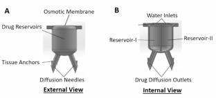
Glioblastoma is a highly malignant brain tumor with limited survival rates due to challenges in complete surgical excision, high recurrence (> 90%), and the inefficacy of systemic drug delivery. Significant efforts have been made to develop drug-loaded brain implants, catheters, and wafers aimed at enhancing survival rates by suppressing tumor recurrence. However, these devices often fail due to clogging, reflux, and the inability to be fully implanted intracranially. Furthermore, a lack of tissue penetration, diffusion distance, and duration of therapy have limited effectiveness of these implants. To address existing challenges, this study reports an osmosis-driven, 3D-printed brain implant with the potential for precise device customization to meet therapeutic needs, while negating systemic toxicity. It is capable of being loaded with two distinct therapeutic agents and implanted directly into the tumor resection cavity during surgery. The device features dual reservoirs, osmotic membranes, and precision-engineered needles for anchoring the device in the resection cavity and perfusing. Further, the device was characterized in vitro using 0.2% agarose gel as a brain tissue analog, with food dye as a drug analog and sodium chloride serving as an osmogen. A design of experiment approach was implemented to investigate various parameters, including membrane pore size, osmogen concentration, needle length, and their effects on release rates. The results demonstrated that the optimized implant achieves flow rates of 2.5 ± 0.1 µl/Hr and diffusion distance of up to 15.5 ± 0.4 mm, using 25 nm pore osmotic membranes with 25.3% osmogen concentration, aligning with model predictions.
期刊介绍:
Biomedical Microdevices: BioMEMS and Biomedical Nanotechnology is an interdisciplinary periodical devoted to all aspects of research in the medical diagnostic and therapeutic applications of Micro-Electro-Mechanical Systems (BioMEMS) and nanotechnology for medicine and biology.
General subjects of interest include the design, characterization, testing, modeling and clinical validation of microfabricated systems, and their integration on-chip and in larger functional units. The specific interests of the Journal include systems for neural stimulation and recording, bioseparation technologies such as nanofilters and electrophoretic equipment, miniaturized analytic and DNA identification systems, biosensors, and micro/nanotechnologies for cell and tissue research, tissue engineering, cell transplantation, and the controlled release of drugs and biological molecules.
Contributions reporting on fundamental and applied investigations of the material science, biochemistry, and physics of biomedical microdevices and nanotechnology are encouraged. A non-exhaustive list of fields of interest includes: nanoparticle synthesis, characterization, and validation of therapeutic or imaging efficacy in animal models; biocompatibility; biochemical modification of microfabricated devices, with reference to non-specific protein adsorption, and the active immobilization and patterning of proteins on micro/nanofabricated surfaces; the dynamics of fluids in micro-and-nano-fabricated channels; the electromechanical and structural response of micro/nanofabricated systems; the interactions of microdevices with cells and tissues, including biocompatibility and biodegradation studies; variations in the characteristics of the systems as a function of the micro/nanofabrication parameters.
 求助内容:
求助内容: 应助结果提醒方式:


