{"title":"Diagnosis of cardiac sarcoidosis using glucose metabolic rate from four-dimensional 18F-FDG PET/CT","authors":"Yurie Shirai, Michinobu Nagao, Akihiro Inoue, Atsushi Yamamoto, Koichiro Kaneko, Akiko Sakai, Atsushi Suzuki, Junichi Yamaguchi, Shuji Sakai","doi":"10.1007/s12149-025-02070-3","DOIUrl":null,"url":null,"abstract":"<div><h3>Objective</h3><p><sup>18</sup>F-Fludeoxyglucose (FDG) PET/CT is an effective tool for detecting active cardiac sarcoidosis (CS), but often has difficulty distinguishing CS lesions from physiological myocardial accumulation. We investigated the potential of the glucose metabolic rate (MR<sub>glc</sub>, mg/min/100mL) from four-dimensional FDG PET/CT in distinguishing between CS and physiological accumulation. Additionally, we compared CS delineation between MR<sub>glc</sub> and standardized uptake value (SUV).</p><h3>Methods</h3><p>A total of 192 individuals with CS or suspected CS who underwent four-dimensional FDG PET/CT after 18 h fasting was enrolled. Ultimately, 45 individuals with CS and 14 patients with physiological accumulation, with SUV<sub>mean</sub> ≥ 2.7 accumulation in the left ventricular myocardium, were analyzed. The SUV, MR<sub>glc</sub>, and the ratio of MR<sub>glc</sub> to SUV (MR<sub>glc</sub>/SUV) were calculated for each lesion with SUV<sub>mean</sub> ≥ 2.7 using data acquired between 30 and 50 min on four-dimensional FDG PET/CT. In the CS group, lesion-to-normal myocardium contrast ratios on MR<sub>glc</sub> and SUV images were compared.</p><h3>Results</h3><p>A total of 127 lesions from 45 individuals with CS and 43 physiological accumulations from 14 individuals were analyzed. The SUV, MR<sub>glc,</sub> and MR<sub>glc</sub>/SUV for CS lesions were significantly lower than those for physiological accumulations (SUV, 4.26±1.35 vs. 6.06±3.28; MR<sub>glc</sub>, 1.91 ± 1.02 vs. 3.78 ± 2.11; MR<sub>glc</sub>/SUV, 0.43 ± 0.14 vs. 0.63 ± 0.14; p < 0.0001). Receiver operating characteristic analysis revealed that the ability to discriminate CS lesions from physiological accumulations yielded areas under the curves of 0.656, 0.808, and 0.849; sensitivities of 68, 76, and 73%; and specificities of 61, 72, and 84%, for SUV 4.525, MR<sub>glc</sub> 2.41, and MR<sub>glc</sub>/SUV 0.518. In the CS group, the contrast ratio of lesions was significantly greater on MR<sub>glc</sub> images than on SUV images (6.36 ± 6.17 vs. 2.54 ± 1.03, p < 0.0001).</p><h3>Conclusions</h3><p>MR<sub>glc</sub> from four-dimensional FDG PET/CT is a useful quantitative measure to distinguish CS lesions from physiological accumulation and enables better visualization of CS lesion contrast than SUV.</p></div>","PeriodicalId":8007,"journal":{"name":"Annals of Nuclear Medicine","volume":"39 10","pages":"1103 - 1112"},"PeriodicalIF":2.5000,"publicationDate":"2025-06-14","publicationTypes":"Journal Article","fieldsOfStudy":null,"isOpenAccess":false,"openAccessPdf":"","citationCount":"0","resultStr":null,"platform":"Semanticscholar","paperid":null,"PeriodicalName":"Annals of Nuclear Medicine","FirstCategoryId":"3","ListUrlMain":"https://link.springer.com/article/10.1007/s12149-025-02070-3","RegionNum":4,"RegionCategory":"医学","ArticlePicture":[],"TitleCN":null,"AbstractTextCN":null,"PMCID":null,"EPubDate":"","PubModel":"","JCR":"Q2","JCRName":"RADIOLOGY, NUCLEAR MEDICINE & MEDICAL IMAGING","Score":null,"Total":0}
引用次数: 0
Abstract
Objective
18F-Fludeoxyglucose (FDG) PET/CT is an effective tool for detecting active cardiac sarcoidosis (CS), but often has difficulty distinguishing CS lesions from physiological myocardial accumulation. We investigated the potential of the glucose metabolic rate (MRglc, mg/min/100mL) from four-dimensional FDG PET/CT in distinguishing between CS and physiological accumulation. Additionally, we compared CS delineation between MRglc and standardized uptake value (SUV).
Methods
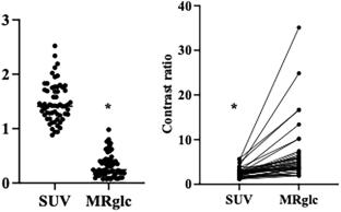
A total of 192 individuals with CS or suspected CS who underwent four-dimensional FDG PET/CT after 18 h fasting was enrolled. Ultimately, 45 individuals with CS and 14 patients with physiological accumulation, with SUVmean ≥ 2.7 accumulation in the left ventricular myocardium, were analyzed. The SUV, MRglc, and the ratio of MRglc to SUV (MRglc/SUV) were calculated for each lesion with SUVmean ≥ 2.7 using data acquired between 30 and 50 min on four-dimensional FDG PET/CT. In the CS group, lesion-to-normal myocardium contrast ratios on MRglc and SUV images were compared.
Results
A total of 127 lesions from 45 individuals with CS and 43 physiological accumulations from 14 individuals were analyzed. The SUV, MRglc, and MRglc/SUV for CS lesions were significantly lower than those for physiological accumulations (SUV, 4.26±1.35 vs. 6.06±3.28; MRglc, 1.91 ± 1.02 vs. 3.78 ± 2.11; MRglc/SUV, 0.43 ± 0.14 vs. 0.63 ± 0.14; p < 0.0001). Receiver operating characteristic analysis revealed that the ability to discriminate CS lesions from physiological accumulations yielded areas under the curves of 0.656, 0.808, and 0.849; sensitivities of 68, 76, and 73%; and specificities of 61, 72, and 84%, for SUV 4.525, MRglc 2.41, and MRglc/SUV 0.518. In the CS group, the contrast ratio of lesions was significantly greater on MRglc images than on SUV images (6.36 ± 6.17 vs. 2.54 ± 1.03, p < 0.0001).
Conclusions
MRglc from four-dimensional FDG PET/CT is a useful quantitative measure to distinguish CS lesions from physiological accumulation and enables better visualization of CS lesion contrast than SUV.
期刊介绍:
Annals of Nuclear Medicine is an official journal of the Japanese Society of Nuclear Medicine. It develops the appropriate application of radioactive substances and stable nuclides in the field of medicine.
The journal promotes the exchange of ideas and information and research in nuclear medicine and includes the medical application of radionuclides and related subjects. It presents original articles, short communications, reviews and letters to the editor.

 求助内容:
求助内容: 应助结果提醒方式:


