Determinants driving the evolution of new multiple sclerosis lesions into chronic active or remyelinated states
IF 3.6
2区 医学
Q2 NEUROIMAGING
引用次数: 0
Abstract
Introduction
Once formed, focal lesions that develop in patients with multiple sclerosis (MS) can follow different trajectories. We aimed at identifying early clinical and MRI features associated with the evolution of new MS lesions into chronic-active versus remyelinated states.
Methods
New contrast-enhancing (CE) lesions were classified after a 12-month follow-up with quantitative susceptibility mapping (QSM) into paramagnetic rim lesions (PRLs, representing chronic-active lesions) and isointense QSM lesions (ISO, representing remyelinated lesions). SHapley Additive exPlanations (SHAP) analysis, which highlights the most relevant features contributing to model predictions, was conducted using baseline clinical and MRI characteristics. A risk score was calculated for PRL and ISO classifications using the four most influential features for each task.
Results
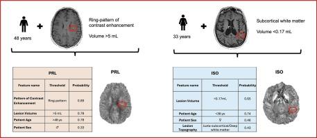
A total of 111 lesions from 44 MS patients were analyzed. At 12 months, 13 % lesions were classified as PRL and 45 % as ISO. The key predictive features were similar for both classes (lesion volume, patient age and sex) except for the pattern of contrast enhancement (which was selected for PRL classification) and lesion topography (which was selected for ISO classification). Older age (>48 years), male sex, bigger lesion volume (>5 mL) and the presence of a ring pattern of contrast enhancement favored PRLs, while younger age (<36 years), female sex, smaller lesion volume (<0.17 mL) and the juxta-subcortical/deep white matter location favored ISO.
Interpretation
The outcome of a new MS lesion can be predicted at lesion onset considering few clinically accessible features.
驱动新发多发性硬化症病变演变为慢性活动性或再髓化状态的决定因素
在多发性硬化症(MS)患者中,局灶性病变一旦形成,可能会有不同的发展轨迹。我们的目的是确定早期临床和MRI特征与新发MS病变演变为慢性活动性和再髓鞘化状态相关。方法经12个月的定量敏感性定位(QSM)随访,将新发对比增强(CE)病变分为顺磁边缘病变(prl,代表慢性活动性病变)和等强度对比增强病变(ISO,代表髓鞘再生病变)。SHapley加性解释(SHAP)分析强调了与模型预测最相关的特征,使用基线临床和MRI特征进行。使用每个任务的四个最具影响力的特征计算PRL和ISO分类的风险评分。结果共分析44例MS患者的111个病变。12个月时,13%的病变被分类为PRL, 45%的病变被分类为ISO。除了对比增强模式(用于PRL分类)和病变地形(用于ISO分类)外,两类的关键预测特征(病变体积、患者年龄和性别)相似。年龄较大(48岁)、男性、病变体积较大(5ml)、有环形对比增强倾向于prl,而年龄较小(36岁)、女性、病变体积较小(0.17 mL)、皮层下近/深部白质位置倾向于ISO。解释:考虑到一些临床可获得的特征,新的MS病变的结果可以在病变开始时预测。
本文章由计算机程序翻译,如有差异,请以英文原文为准。
求助全文
约1分钟内获得全文
求助全文
来源期刊

Neuroimage-Clinical
NEUROIMAGING-
CiteScore
7.50
自引率
4.80%
发文量
368
审稿时长
52 days
期刊介绍:
NeuroImage: Clinical, a journal of diseases, disorders and syndromes involving the Nervous System, provides a vehicle for communicating important advances in the study of abnormal structure-function relationships of the human nervous system based on imaging.
The focus of NeuroImage: Clinical is on defining changes to the brain associated with primary neurologic and psychiatric diseases and disorders of the nervous system as well as behavioral syndromes and developmental conditions. The main criterion for judging papers is the extent of scientific advancement in the understanding of the pathophysiologic mechanisms of diseases and disorders, in identification of functional models that link clinical signs and symptoms with brain function and in the creation of image based tools applicable to a broad range of clinical needs including diagnosis, monitoring and tracking of illness, predicting therapeutic response and development of new treatments. Papers dealing with structure and function in animal models will also be considered if they reveal mechanisms that can be readily translated to human conditions.
 求助内容:
求助内容: 应助结果提醒方式:


