Liam O Cunningham, Aravinda Ganapathy, Cihat Eldeniz, Jeffery A Weisman, Kevin E Lindsay, Udayabhanu Jammalamadaka, Karthik Tappa, Amber Salter, Hongyu An, Pamela K Woodard, David H Ballard
{"title":"3D printed vitamin D impregnated catheters for magnetic resonance-guided interventions: proof of concept and imaging characteristics.","authors":"Liam O Cunningham, Aravinda Ganapathy, Cihat Eldeniz, Jeffery A Weisman, Kevin E Lindsay, Udayabhanu Jammalamadaka, Karthik Tappa, Amber Salter, Hongyu An, Pamela K Woodard, David H Ballard","doi":"10.1186/s41205-025-00273-y","DOIUrl":null,"url":null,"abstract":"<p><strong>Background: </strong>Catheters used for magnetic resonance (MR)-guided interventions require intra-catheter coils and often produce artifacts. This study aimed to fabricate 3D-printed catheters impregnated with vitamin D solution to allow for optimal visualization during MR-guided procedures.</p><p><strong>Methods: </strong>3D printing was used to fabricate catheters impregnated with vitamin D solution. Computer-aided design files were generated for a size 18 French catheter prototype with a compartment for vitamin D solution to be manually introduced into the catheter's lumen and sealed via thermoplastic welding. Polylactic acid (PLA) bioplastic was 3D printed into filaments via material extrusion (FDM<sup>®</sup>, Stratasys, Eden Prairie, MN) on a 5th generation Replicator 3D printer (MakerBot). Three different forms of vitamin D were used, cholecalciferol, ergocalciferol, and calcitriol, and 0.9% normal saline served as a control. Three prints of each catheter type were fabricated and scanned using a 1.5 T MR whole body scanner (Avanto, Siemens Healthcare) inside a small flex loop surface radiofrequency (RF) coil. A 3D gradient recalled echo (GRE) sequence was used with the following acquisition parameters: 4.52/11 ms TE/TR, 15° flip angle, 256 × 256 matrix with 0.5 mm × 0.5 mm in-plane resolution, 24 coronal slabs, 2 mm thickness, and 140 Hz receiver bandwidth. Three averages were used to improve the signal-to-noise ratio (SNR). The GRE sequence was run with 4 different flip angles: 3°, 15°, 30°, and 45° to perform T1 mapping.</p><p><strong>Results: </strong>All 3D-printed catheters impregnated with vitamin D produced a signal on MR. SNR for vitamin D catheters was similar across the various forms of vitamin D: mean SNRs for 100% cholecalciferol, ergocalciferol, and calcitriol were 138, 139, and 130. Mean SNR and contrast-to-noise ratio (CNR) for vitamin D catheters were significantly higher than the control saline catheter (p < 0.001, for both SNR and CNR). T1 values were lower in vitamin D-impregnated catheters compared to the saline control (228 ± 67 ms and 3371 ± 493 ms, respectively; p < 0.0001), indicating a better signal.</p><p><strong>Conclusions: </strong>3D printing of catheters impregnated with vitamin D is feasible and can potentially optimize MR-guided procedures.</p>","PeriodicalId":72036,"journal":{"name":"3D printing in medicine","volume":"11 1","pages":"27"},"PeriodicalIF":3.1000,"publicationDate":"2025-06-13","publicationTypes":"Journal Article","fieldsOfStudy":null,"isOpenAccess":false,"openAccessPdf":"https://www.ncbi.nlm.nih.gov/pmc/articles/PMC12164080/pdf/","citationCount":"0","resultStr":null,"platform":"Semanticscholar","paperid":null,"PeriodicalName":"3D printing in medicine","FirstCategoryId":"1085","ListUrlMain":"https://doi.org/10.1186/s41205-025-00273-y","RegionNum":0,"RegionCategory":null,"ArticlePicture":[],"TitleCN":null,"AbstractTextCN":null,"PMCID":null,"EPubDate":"","PubModel":"","JCR":"Q1","JCRName":"RADIOLOGY, NUCLEAR MEDICINE & MEDICAL IMAGING","Score":null,"Total":0}
引用次数: 0
Abstract
Background: Catheters used for magnetic resonance (MR)-guided interventions require intra-catheter coils and often produce artifacts. This study aimed to fabricate 3D-printed catheters impregnated with vitamin D solution to allow for optimal visualization during MR-guided procedures.
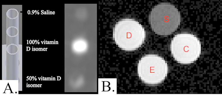
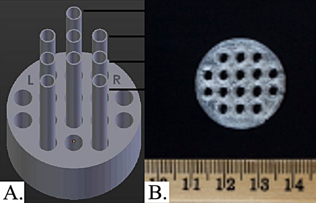
Methods: 3D printing was used to fabricate catheters impregnated with vitamin D solution. Computer-aided design files were generated for a size 18 French catheter prototype with a compartment for vitamin D solution to be manually introduced into the catheter's lumen and sealed via thermoplastic welding. Polylactic acid (PLA) bioplastic was 3D printed into filaments via material extrusion (FDM®, Stratasys, Eden Prairie, MN) on a 5th generation Replicator 3D printer (MakerBot). Three different forms of vitamin D were used, cholecalciferol, ergocalciferol, and calcitriol, and 0.9% normal saline served as a control. Three prints of each catheter type were fabricated and scanned using a 1.5 T MR whole body scanner (Avanto, Siemens Healthcare) inside a small flex loop surface radiofrequency (RF) coil. A 3D gradient recalled echo (GRE) sequence was used with the following acquisition parameters: 4.52/11 ms TE/TR, 15° flip angle, 256 × 256 matrix with 0.5 mm × 0.5 mm in-plane resolution, 24 coronal slabs, 2 mm thickness, and 140 Hz receiver bandwidth. Three averages were used to improve the signal-to-noise ratio (SNR). The GRE sequence was run with 4 different flip angles: 3°, 15°, 30°, and 45° to perform T1 mapping.
Results: All 3D-printed catheters impregnated with vitamin D produced a signal on MR. SNR for vitamin D catheters was similar across the various forms of vitamin D: mean SNRs for 100% cholecalciferol, ergocalciferol, and calcitriol were 138, 139, and 130. Mean SNR and contrast-to-noise ratio (CNR) for vitamin D catheters were significantly higher than the control saline catheter (p < 0.001, for both SNR and CNR). T1 values were lower in vitamin D-impregnated catheters compared to the saline control (228 ± 67 ms and 3371 ± 493 ms, respectively; p < 0.0001), indicating a better signal.
Conclusions: 3D printing of catheters impregnated with vitamin D is feasible and can potentially optimize MR-guided procedures.


 求助内容:
求助内容: 应助结果提醒方式:


