Fariztah Sukainah Nur Fathimah, Sauli Ari Widjaja, Wimbo Sasono, Ima Yustiarini, Muhammad Firmansjah, Ady Dwi Prakosa, Aulia Kezia Mulyazhara, Soebagijo Adi Soelistijo
{"title":"Retinal vessel tortuosity and fractal dimension in diabetic retinopathy.","authors":"Fariztah Sukainah Nur Fathimah, Sauli Ari Widjaja, Wimbo Sasono, Ima Yustiarini, Muhammad Firmansjah, Ady Dwi Prakosa, Aulia Kezia Mulyazhara, Soebagijo Adi Soelistijo","doi":"10.1186/s40942-025-00688-z","DOIUrl":null,"url":null,"abstract":"<p><strong>Background: </strong>Retinal vessel geometry characteristic have been studied as one of the signs of microvascular changes in diabetic retinopathy (DR) that necessitates early screening. This study aimed to investigate the differences in retinal vessel tortuosity (VT) and fractal dimension (FD) between patients with and without DR.</p><p><strong>Methods: </strong>This retrospective study analyzed medical records and OCT-A images of DR and No-DR patients. DR severity was graded by a vitreoretinal specialist following the International Clinical Diabetic Retinopathy and Diabetic Macular Edema Severity Scales. Retinal VT and FD were quantified using ImageJ software. Comparison between groups using non-parametric and Generalized Estimating Equations (GEE) statistical analysis combined with cluster bootstrapping.</p><p><strong>Results: </strong>We analyzed 96 (161 eyes) with the mean age of 52.7 ± 9.9 years. Compared to No-DR, VT was significantly higher in all DR groups (p < 0.05). Mild non proliferative DR (β = +0.0621), Moderate NPDR (β = +0.0412), Severe NPDR (β = +0.0441), and proliferative DR (β = +0.0404). FD of the superficial capillary plexus (SCP) showed no significant difference among the groups and a significantly lower FD of the deep capillary plexus (DCP) compared to the No-DR groups (moderate NPDR (β = -0.0131), severe NPDR ( β = -0.0316) and PDR ( β = -0.0326)).</p><p><strong>Conclusion: </strong>Compared to No-DR group, VT was found significantly higher in DR group, and FD of the DCP found significantly lower in the DR group. These parameters offer unique insights beyond simple vessel loss and complementary information into the geometric complexity and structural alterations of the retinal microvasculature in DR.</p>","PeriodicalId":14289,"journal":{"name":"International Journal of Retina and Vitreous","volume":"11 1","pages":"64"},"PeriodicalIF":2.4000,"publicationDate":"2025-06-12","publicationTypes":"Journal Article","fieldsOfStudy":null,"isOpenAccess":false,"openAccessPdf":"https://www.ncbi.nlm.nih.gov/pmc/articles/PMC12164056/pdf/","citationCount":"0","resultStr":null,"platform":"Semanticscholar","paperid":null,"PeriodicalName":"International Journal of Retina and Vitreous","FirstCategoryId":"1085","ListUrlMain":"https://doi.org/10.1186/s40942-025-00688-z","RegionNum":0,"RegionCategory":null,"ArticlePicture":[],"TitleCN":null,"AbstractTextCN":null,"PMCID":null,"EPubDate":"","PubModel":"","JCR":"Q2","JCRName":"OPHTHALMOLOGY","Score":null,"Total":0}
引用次数: 0
Abstract
Background: Retinal vessel geometry characteristic have been studied as one of the signs of microvascular changes in diabetic retinopathy (DR) that necessitates early screening. This study aimed to investigate the differences in retinal vessel tortuosity (VT) and fractal dimension (FD) between patients with and without DR.
Methods: This retrospective study analyzed medical records and OCT-A images of DR and No-DR patients. DR severity was graded by a vitreoretinal specialist following the International Clinical Diabetic Retinopathy and Diabetic Macular Edema Severity Scales. Retinal VT and FD were quantified using ImageJ software. Comparison between groups using non-parametric and Generalized Estimating Equations (GEE) statistical analysis combined with cluster bootstrapping.
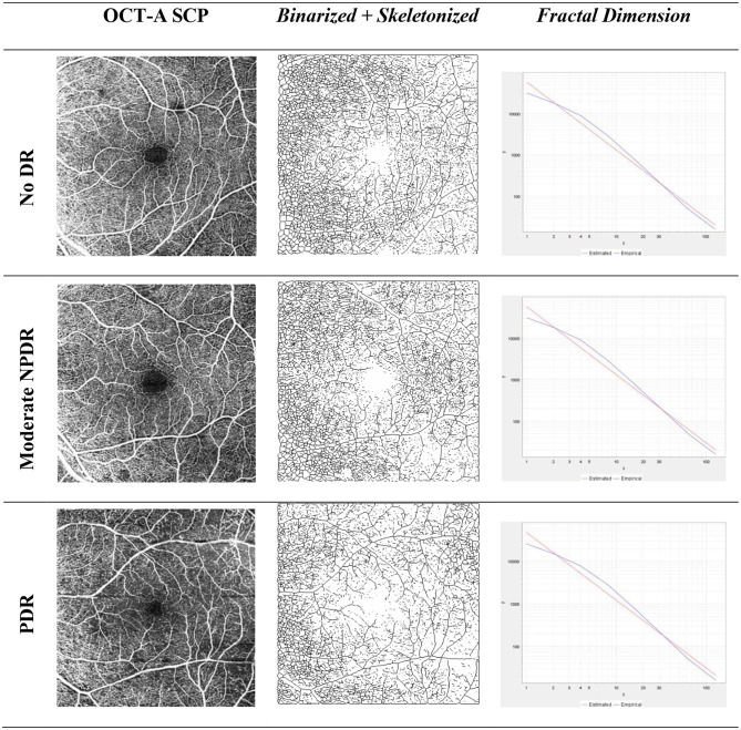
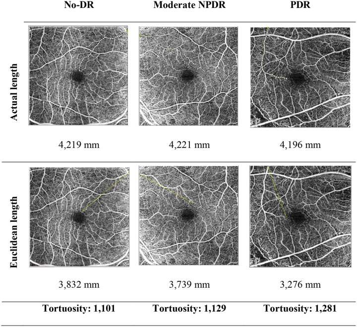
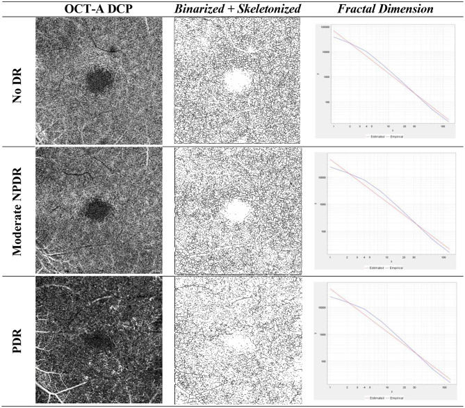
Results: We analyzed 96 (161 eyes) with the mean age of 52.7 ± 9.9 years. Compared to No-DR, VT was significantly higher in all DR groups (p < 0.05). Mild non proliferative DR (β = +0.0621), Moderate NPDR (β = +0.0412), Severe NPDR (β = +0.0441), and proliferative DR (β = +0.0404). FD of the superficial capillary plexus (SCP) showed no significant difference among the groups and a significantly lower FD of the deep capillary plexus (DCP) compared to the No-DR groups (moderate NPDR (β = -0.0131), severe NPDR ( β = -0.0316) and PDR ( β = -0.0326)).
Conclusion: Compared to No-DR group, VT was found significantly higher in DR group, and FD of the DCP found significantly lower in the DR group. These parameters offer unique insights beyond simple vessel loss and complementary information into the geometric complexity and structural alterations of the retinal microvasculature in DR.
期刊介绍:
International Journal of Retina and Vitreous focuses on the ophthalmic subspecialty of vitreoretinal disorders. The journal presents original articles on new approaches to diagnosis, outcomes of clinical trials, innovations in pharmacological therapy and surgical techniques, as well as basic science advances that impact clinical practice. Topical areas include, but are not limited to: -Imaging of the retina, choroid and vitreous -Innovations in optical coherence tomography (OCT) -Small-gauge vitrectomy, retinal detachment, chromovitrectomy -Electroretinography (ERG), microperimetry, other functional tests -Intraocular tumors -Retinal pharmacotherapy & drug delivery -Diabetic retinopathy & other vascular diseases -Age-related macular degeneration (AMD) & other macular entities

 求助内容:
求助内容: 应助结果提醒方式:


