{"title":"[Gastric Undifferentiated Pleomorphic Sarcoma Presenting as a Rapidly Growing Subepithelial Tumor].","authors":"Dae Gon Ryu, Cheol Woong Choi","doi":"10.7704/kjhugr.2024.0033","DOIUrl":null,"url":null,"abstract":"<p><p>A 48-year-old man presented to our hospital for evaluation of melena. Abdominal computed tomography (CT) revealed an endophytic contrast-enhanced mass (approximately 9 cm) in the gastric fundus and upper body. CT showed no evidence of metastasis to other organs. Endoscopic examination revealed a large mass accompanied by an exudate and signs of surface hemorrhage. The shape of the mass resembled that of a subepithelial tumor, and >50% of the mucosa was peeled off. Endoscopic ultrasonography revealed hyperechoic spreading and dotor rod-shaped anechoic lesions arranged within an overall hypoechoic mass. The patient underwent total gastrectomy with lymph node dissection and was diagnosed with undifferentiated pleomorphic sarcoma without lymph node metastasis. Undifferentiated pleomorphic sarcoma, a high-grade aggressive soft tissue sarcoma, predominantly involves the extremities and rarely the stomach. It is usually asymptomatic but tends to grow rapidly. Endoscopic imaging performed a year prior to presentation was unremarkable. We report a case of a gastric undifferentiated pleomorphic sarcoma that presented as a rapidly growing subepithelial tumor. Malignancies other than a gastrointestinal stromal tumor should be considered in the differential diagnosis in patients presenting with a rapidly growing gastric subepithelial tumor.</p>","PeriodicalId":520887,"journal":{"name":"The Korean journal of helicobacter and upper gastrointestinal research","volume":"24 3","pages":"276-280"},"PeriodicalIF":0.0000,"publicationDate":"2024-09-01","publicationTypes":"Journal Article","fieldsOfStudy":null,"isOpenAccess":false,"openAccessPdf":"https://www.ncbi.nlm.nih.gov/pmc/articles/PMC11967536/pdf/","citationCount":"0","resultStr":null,"platform":"Semanticscholar","paperid":null,"PeriodicalName":"The Korean journal of helicobacter and upper gastrointestinal research","FirstCategoryId":"1085","ListUrlMain":"https://doi.org/10.7704/kjhugr.2024.0033","RegionNum":0,"RegionCategory":null,"ArticlePicture":[],"TitleCN":null,"AbstractTextCN":null,"PMCID":null,"EPubDate":"2024/9/9 0:00:00","PubModel":"Epub","JCR":"","JCRName":"","Score":null,"Total":0}
引用次数: 0
Abstract
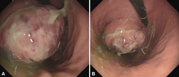
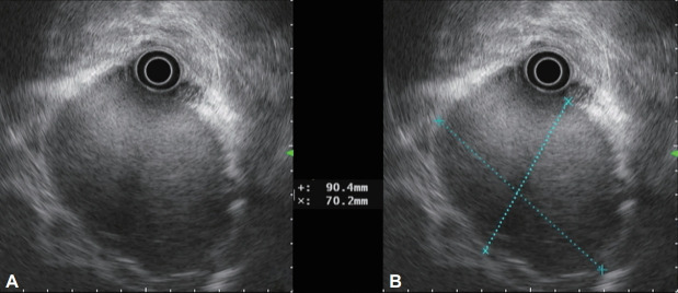
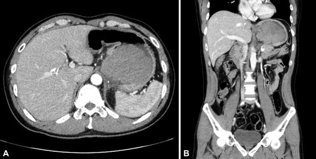
A 48-year-old man presented to our hospital for evaluation of melena. Abdominal computed tomography (CT) revealed an endophytic contrast-enhanced mass (approximately 9 cm) in the gastric fundus and upper body. CT showed no evidence of metastasis to other organs. Endoscopic examination revealed a large mass accompanied by an exudate and signs of surface hemorrhage. The shape of the mass resembled that of a subepithelial tumor, and >50% of the mucosa was peeled off. Endoscopic ultrasonography revealed hyperechoic spreading and dotor rod-shaped anechoic lesions arranged within an overall hypoechoic mass. The patient underwent total gastrectomy with lymph node dissection and was diagnosed with undifferentiated pleomorphic sarcoma without lymph node metastasis. Undifferentiated pleomorphic sarcoma, a high-grade aggressive soft tissue sarcoma, predominantly involves the extremities and rarely the stomach. It is usually asymptomatic but tends to grow rapidly. Endoscopic imaging performed a year prior to presentation was unremarkable. We report a case of a gastric undifferentiated pleomorphic sarcoma that presented as a rapidly growing subepithelial tumor. Malignancies other than a gastrointestinal stromal tumor should be considered in the differential diagnosis in patients presenting with a rapidly growing gastric subepithelial tumor.

 求助内容:
求助内容: 应助结果提醒方式:


