Richard Hall, Cassandra Bruce-Brand, Washington Mudini, Alessandro Pietro Aldera
{"title":"Role of <i>Helicobacter pylori</i> Immunohistochemistry in the Histopathological Assessment of Inflamed Endoscopic Gastric Biopsies.","authors":"Richard Hall, Cassandra Bruce-Brand, Washington Mudini, Alessandro Pietro Aldera","doi":"10.7704/kjhugr.2023.0048","DOIUrl":null,"url":null,"abstract":"<p><strong>Objectives: </strong>The identification of <i>Helicobacter pylori</i> is one of the main tasks of diagnostic histopathologists when evaluating endoscopic gastric biopsies. The sensitivity and specificity of different stains that facilitate this identification vary. Despite the existing guidelines, many histopathology laboratories perform routine histochemical staining of all gastric biopsies to improve turnaround times. This study assessed the utility of an <i>H. pylori</i> immunohistochemical (IHC) stain compared with a routinely used histochemical stain, cresyl violet (CV), in the South African setting.</p><p><strong>Methods: </strong>Cases were identified retrospectively, and original histopathology reports were used to establish the \"ground truth\" diagnoses. Three pathologists independently evaluated the CV and IHC stains; each pathologist was timed in a standardized manner. The sensitivity, specificity, interobserver variability, and time taken to identify <i>H. pylori</i> with each stain were compared.</p><p><strong>Results: </strong>The overall sensitivity and specificity for IHC staining (85.2% and 97.7%, respectively) were higher than those for CV staining (64.5% and 90.6%, respectively). Detection of <i>H. pylori</i> took an average of 16 and 49 seconds using the IHC and CV stains, respectively. The prevalence of <i>H. pylori</i> in our laboratory was 23.7%, which is lower than the reported national prevalence in South Africa.</p><p><strong>Conclusions: </strong>IHC stain-based detection of <i>H. pylori</i> in inflamed gastric biopsies demonstrated superior sensitivity and specificity than CV staining. This was particularly true for cases involving patients with low bacterial loads. The interpretation of <i>H. pylori</i> IHC staining is much faster than that associated with CV staining, which is important in centers with high caseloads and shortages of pathologists.</p>","PeriodicalId":520887,"journal":{"name":"The Korean journal of helicobacter and upper gastrointestinal research","volume":"24 1","pages":"45-51"},"PeriodicalIF":0.0000,"publicationDate":"2024-03-01","publicationTypes":"Journal Article","fieldsOfStudy":null,"isOpenAccess":false,"openAccessPdf":"https://www.ncbi.nlm.nih.gov/pmc/articles/PMC11967533/pdf/","citationCount":"0","resultStr":null,"platform":"Semanticscholar","paperid":null,"PeriodicalName":"The Korean journal of helicobacter and upper gastrointestinal research","FirstCategoryId":"1085","ListUrlMain":"https://doi.org/10.7704/kjhugr.2023.0048","RegionNum":0,"RegionCategory":null,"ArticlePicture":[],"TitleCN":null,"AbstractTextCN":null,"PMCID":null,"EPubDate":"2024/3/8 0:00:00","PubModel":"Epub","JCR":"","JCRName":"","Score":null,"Total":0}
引用次数: 0
Abstract
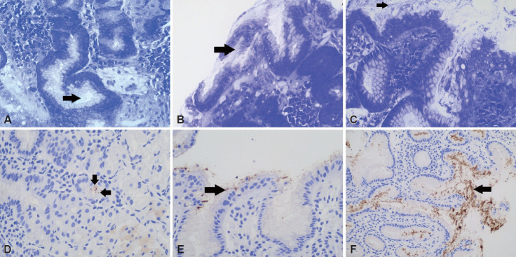
Objectives: The identification of Helicobacter pylori is one of the main tasks of diagnostic histopathologists when evaluating endoscopic gastric biopsies. The sensitivity and specificity of different stains that facilitate this identification vary. Despite the existing guidelines, many histopathology laboratories perform routine histochemical staining of all gastric biopsies to improve turnaround times. This study assessed the utility of an H. pylori immunohistochemical (IHC) stain compared with a routinely used histochemical stain, cresyl violet (CV), in the South African setting.
Methods: Cases were identified retrospectively, and original histopathology reports were used to establish the "ground truth" diagnoses. Three pathologists independently evaluated the CV and IHC stains; each pathologist was timed in a standardized manner. The sensitivity, specificity, interobserver variability, and time taken to identify H. pylori with each stain were compared.
Results: The overall sensitivity and specificity for IHC staining (85.2% and 97.7%, respectively) were higher than those for CV staining (64.5% and 90.6%, respectively). Detection of H. pylori took an average of 16 and 49 seconds using the IHC and CV stains, respectively. The prevalence of H. pylori in our laboratory was 23.7%, which is lower than the reported national prevalence in South Africa.
Conclusions: IHC stain-based detection of H. pylori in inflamed gastric biopsies demonstrated superior sensitivity and specificity than CV staining. This was particularly true for cases involving patients with low bacterial loads. The interpretation of H. pylori IHC staining is much faster than that associated with CV staining, which is important in centers with high caseloads and shortages of pathologists.

 求助内容:
求助内容: 应助结果提醒方式:


