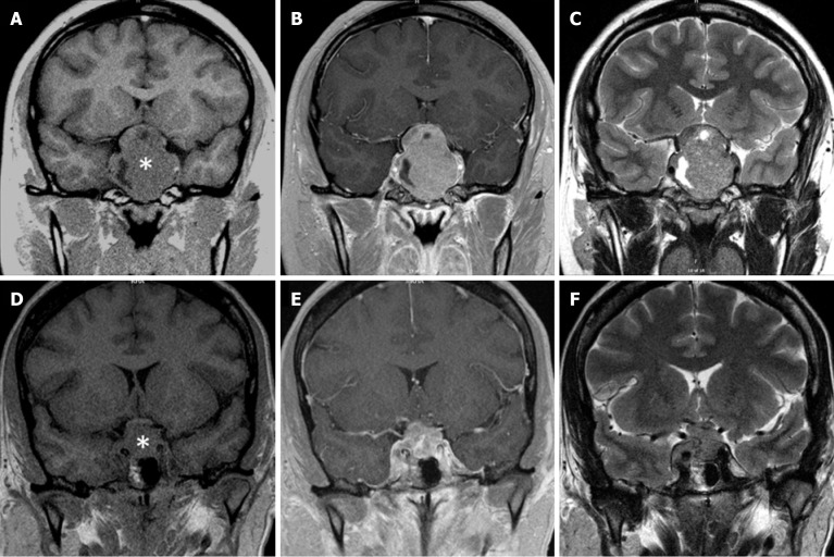
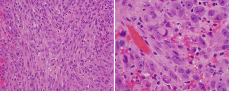
{"title":"Specific imaging features of sellar atypical teratoid/rhabdoid tumor or the lack of thereof.","authors":"Run Yu","doi":"10.4329/wjr.v17.i5.106975","DOIUrl":null,"url":null,"abstract":"<p><p>Primary sellar atypical teratoid/rhabdoid tumor (AT/RT) is the most aggressive sellar mass. Although rare, sellar AT/RT exhibits a very relentless clinical course and usually results in death within months to a few years after diagnosis. The best clinical evidence suggests that surgical debulking and timely adjuvant chemoradiation are most effective in prolonging survival. A preoperative radiological diagnosis of sellar AT/RT thus is crucial in informing patients and physicians about this devastating disease. This minireview summaries the imaging features of sellar AT/RT. magnetic resonance imaging features of sellar AT/RT and the much more common sellar mass, pituitary macroadenoma, are similar in most aspects: They are both isointense to brain gray matter on T1 and T2 imaging and enhance upon gadolinium administration. Suprasellar extension and cavernous sinus invasion are present in practically all cases of sellar AT/RT, but are also present in 50%-75% of pituitary macroadenomas, especially in large ones, suggesting that suprasellar extension and cavernous sinus invasion disproportionate to the tumor size may favor sellar AT/RT diagnosis. Since sellar AT/RT grows very rapidly and does not allow significant remodeling of perisellar structures, the imaging features of perisellar structures such as optic chiasm and cavernous sinus may be key for imaging diagnosis of sellar AT/RT although they have not been well described in sellar AT/RT. In limited cases of sellar AT/RT, optic chiasm degeneration and thinning, which are very common in pituitary macroadenoma, are not present, giving hope for using features of perisellar structures to diagnose sellar AT/RT by imaging.</p>","PeriodicalId":23819,"journal":{"name":"World journal of radiology","volume":"17 5","pages":"106975"},"PeriodicalIF":1.5000,"publicationDate":"2025-05-28","publicationTypes":"Journal Article","fieldsOfStudy":null,"isOpenAccess":false,"openAccessPdf":"https://www.ncbi.nlm.nih.gov/pmc/articles/PMC12149976/pdf/","citationCount":"0","resultStr":null,"platform":"Semanticscholar","paperid":null,"PeriodicalName":"World journal of radiology","FirstCategoryId":"1085","ListUrlMain":"https://doi.org/10.4329/wjr.v17.i5.106975","RegionNum":0,"RegionCategory":null,"ArticlePicture":[],"TitleCN":null,"AbstractTextCN":null,"PMCID":null,"EPubDate":"","PubModel":"","JCR":"Q3","JCRName":"RADIOLOGY, NUCLEAR MEDICINE & MEDICAL IMAGING","Score":null,"Total":0}
引用次数: 0
Abstract
Primary sellar atypical teratoid/rhabdoid tumor (AT/RT) is the most aggressive sellar mass. Although rare, sellar AT/RT exhibits a very relentless clinical course and usually results in death within months to a few years after diagnosis. The best clinical evidence suggests that surgical debulking and timely adjuvant chemoradiation are most effective in prolonging survival. A preoperative radiological diagnosis of sellar AT/RT thus is crucial in informing patients and physicians about this devastating disease. This minireview summaries the imaging features of sellar AT/RT. magnetic resonance imaging features of sellar AT/RT and the much more common sellar mass, pituitary macroadenoma, are similar in most aspects: They are both isointense to brain gray matter on T1 and T2 imaging and enhance upon gadolinium administration. Suprasellar extension and cavernous sinus invasion are present in practically all cases of sellar AT/RT, but are also present in 50%-75% of pituitary macroadenomas, especially in large ones, suggesting that suprasellar extension and cavernous sinus invasion disproportionate to the tumor size may favor sellar AT/RT diagnosis. Since sellar AT/RT grows very rapidly and does not allow significant remodeling of perisellar structures, the imaging features of perisellar structures such as optic chiasm and cavernous sinus may be key for imaging diagnosis of sellar AT/RT although they have not been well described in sellar AT/RT. In limited cases of sellar AT/RT, optic chiasm degeneration and thinning, which are very common in pituitary macroadenoma, are not present, giving hope for using features of perisellar structures to diagnose sellar AT/RT by imaging.

 求助内容:
求助内容: 应助结果提醒方式:


