Md Mobarak Karim, Ruijiao Sun, Behzad Khajavi, Manmohan Singh, Yogeshwari S Ambekar, Alexander W Schill, Salavat R Aglyamov, David Mayerich, Kirill V Larin
{"title":"Multimodal optical coherence tomography and two-photon light sheet fluorescence microscopy for embryo imaging.","authors":"Md Mobarak Karim, Ruijiao Sun, Behzad Khajavi, Manmohan Singh, Yogeshwari S Ambekar, Alexander W Schill, Salavat R Aglyamov, David Mayerich, Kirill V Larin","doi":"10.1117/1.JBO.30.6.060501","DOIUrl":null,"url":null,"abstract":"<p><strong>Significance: </strong>Structural and molecular imaging of the developing embryo can provide deep insights into the development of various pathologies, but few techniques enable the simultaneous detection of these parameters. We demonstrate the first use of combined optical coherence tomography and two-photon light sheet fluorescence microscopy (2P-LSFM) for simultaneous structural and molecular imaging.</p><p><strong>Aim: </strong>We aim to develop a multimodal high-resolution embryonic system that facilitates simultaneous structural and molecular embryonic imaging.</p><p><strong>Approach: </strong>We have developed a multimodal imaging system in which the optical coherence tomography (OCT) and light sheet illumination beams were optically co-aligned and scanned through the galvanometer-mounted mirrors and the same illumination objective.</p><p><strong>Results: </strong>The swept-source OCT system provides a lateral resolution of <math><mrow><mo>∼</mo> <mn>15</mn> <mtext> </mtext> <mi>μ</mi> <mi>m</mi></mrow> </math> and an axial resolution of <math><mrow><mo>∼</mo> <mn>7</mn> <mtext> </mtext> <mi>μ</mi> <mi>m</mi></mrow> </math> . The 2P-LSFM light sheet thickness was <math><mrow><mo>∼</mo> <mn>10</mn> <mtext> </mtext> <mi>μ</mi> <mi>m</mi></mrow> </math> , and the transverse resolution was <math><mrow><mo>∼</mo> <mn>2</mn> <mtext> </mtext> <mi>μ</mi> <mi>m</mi></mrow> </math> . We have demonstrated the system's capabilities using fluorescent microbeads and fluorescently tagged mouse embryos.</p><p><strong>Conclusions: </strong>The co-alignment of the OCT and 2P-LSFM systems enables simple image registration and high-throughput multimodal imaging.</p>","PeriodicalId":15264,"journal":{"name":"Journal of Biomedical Optics","volume":"30 6","pages":"060501"},"PeriodicalIF":2.9000,"publicationDate":"2025-06-01","publicationTypes":"Journal Article","fieldsOfStudy":null,"isOpenAccess":false,"openAccessPdf":"https://www.ncbi.nlm.nih.gov/pmc/articles/PMC12152587/pdf/","citationCount":"0","resultStr":null,"platform":"Semanticscholar","paperid":null,"PeriodicalName":"Journal of Biomedical Optics","FirstCategoryId":"3","ListUrlMain":"https://doi.org/10.1117/1.JBO.30.6.060501","RegionNum":3,"RegionCategory":"医学","ArticlePicture":[],"TitleCN":null,"AbstractTextCN":null,"PMCID":null,"EPubDate":"2025/6/11 0:00:00","PubModel":"Epub","JCR":"Q2","JCRName":"BIOCHEMICAL RESEARCH METHODS","Score":null,"Total":0}
引用次数: 0
Abstract
Significance: Structural and molecular imaging of the developing embryo can provide deep insights into the development of various pathologies, but few techniques enable the simultaneous detection of these parameters. We demonstrate the first use of combined optical coherence tomography and two-photon light sheet fluorescence microscopy (2P-LSFM) for simultaneous structural and molecular imaging.
Aim: We aim to develop a multimodal high-resolution embryonic system that facilitates simultaneous structural and molecular embryonic imaging.
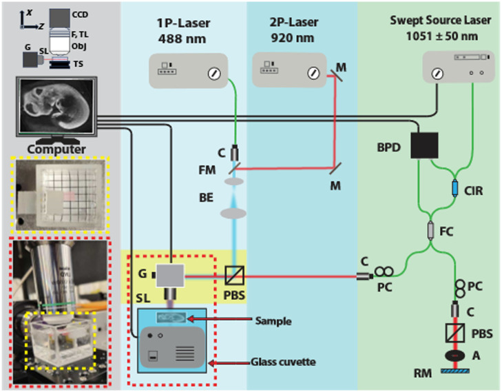
Approach: We have developed a multimodal imaging system in which the optical coherence tomography (OCT) and light sheet illumination beams were optically co-aligned and scanned through the galvanometer-mounted mirrors and the same illumination objective.
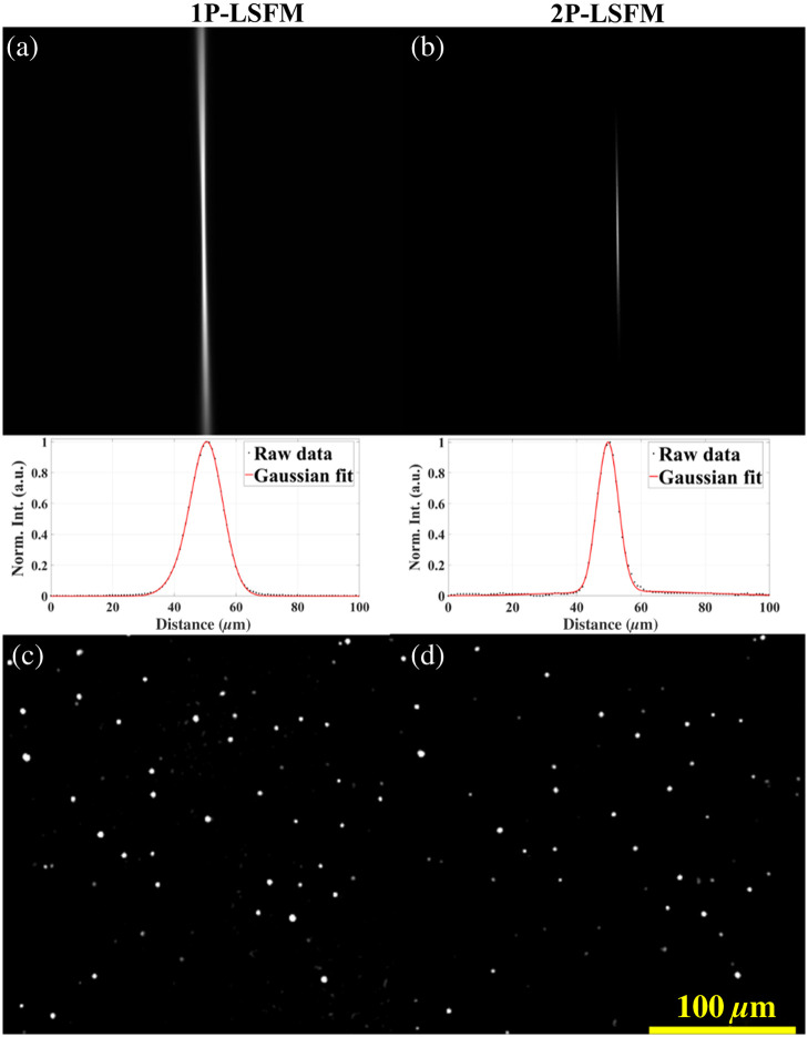
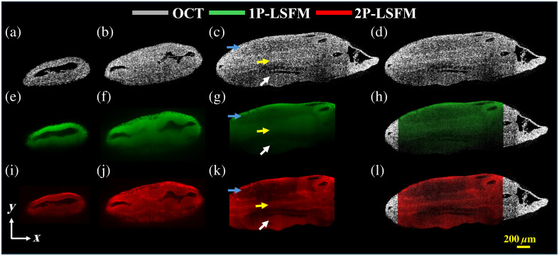
Results: The swept-source OCT system provides a lateral resolution of and an axial resolution of . The 2P-LSFM light sheet thickness was , and the transverse resolution was . We have demonstrated the system's capabilities using fluorescent microbeads and fluorescently tagged mouse embryos.
Conclusions: The co-alignment of the OCT and 2P-LSFM systems enables simple image registration and high-throughput multimodal imaging.
期刊介绍:
The Journal of Biomedical Optics publishes peer-reviewed papers on the use of modern optical technology for improved health care and biomedical research.

 求助内容:
求助内容: 应助结果提醒方式:


