Hemisection versus conventional extraction as interceptive treatment in congenitally missing mandibular second premolars: a randomised controlled split-mouth trial.
Sarah Abdul Jabbar, Shaker Nawaia, Vini Rughwani, Ken Hansen, Julia Naoumova
{"title":"Hemisection versus conventional extraction as interceptive treatment in congenitally missing mandibular second premolars: a randomised controlled split-mouth trial.","authors":"Sarah Abdul Jabbar, Shaker Nawaia, Vini Rughwani, Ken Hansen, Julia Naoumova","doi":"10.1093/ejo/cjaf043","DOIUrl":null,"url":null,"abstract":"<p><strong>Background: </strong>The congenital absence of mandibular second premolars is a common anomaly requiring careful treatment planning. Conventional extraction of the primary molar often causes spontaneous space closure but may lead to mesial tipping of adjacent teeth. Hemisection offers an alternative to control tooth movement and reduce tipping. However, evidence comparing hemisection and conventional extraction, particularly on space closure and tooth angulation, is limited.</p><p><strong>Objectives: </strong>To compare conventional extraction with hemisection of the mandibular primary second molars in terms of space closure, tooth angulation, complications and associated economic implications in patients with congenital absence of mandibular second premolars.</p><p><strong>Trial design: </strong>prospective, randomised longitudinal split-mouth.</p><p><strong>Materials and methods: </strong>Patients with bilateral agenesis of the second mandibular molars and unerupted second molars were included and randomly allocated to either extraction or hemisection on the left or right side of the mandible. Clinical and radiographic examinations were conducted at baseline (T1) and after a mean follow-up period of 4.2 years (T2). Measurements of the residual spaces and tooth angulation of the mandibular first molar and premolar following extraction were blinded assessed on panoramic radiographs and cast models. The number of visits, chair time, social costs, and direct and indirect costs were calculated using cost minimisation analysis.</p><p><strong>Results: </strong>A total of 40 patients (25 boys and 15 girls) with a mean age of 10.03 ± 1.07 years at T1 participated. No patient was lost to follow-up. The residual space between the first permanent molar and the first permanent premolar was 2.04 ± 1.67 mm for hemisection and 2.39 ± 1.86 mm for extraction (p = 0.053). A larger residual space was observed between the first permanent premolar and the canine on the hemisection side (1.80 ± 1.01 mm) than on the extraction side (1.55 ± 0.92 mm), (p = 0.045). No difference was found between the interventions regarding the angulation of the first permanent molar (p = 0.0914) or the angulation of the first permanent premolar (p = 0.7812). Hemisection resulted in significantly more complications (p = 0.0176) and was associated with substantially higher material costs, more chair time and higher indirect costs than conventional extraction (p < 0.0001).</p><p><strong>Conclusion: </strong>Hemisection is not recommended as an interceptive extraction option for patients with congenitally missing mandibular second premolars, as only minimal, clinically irrelevant differences were observed compared with conventional extraction. Moreover, hemisection is associated with increased costs and a higher risk of complications.</p><p><strong>Trial registration: </strong>The trial was registered with https://www.researchweb.org/is/sverige, registration number: 967125.</p>","PeriodicalId":11989,"journal":{"name":"European journal of orthodontics","volume":"47 4","pages":""},"PeriodicalIF":2.7000,"publicationDate":"2025-06-12","publicationTypes":"Journal Article","fieldsOfStudy":null,"isOpenAccess":false,"openAccessPdf":"https://www.ncbi.nlm.nih.gov/pmc/articles/PMC12159412/pdf/","citationCount":"0","resultStr":null,"platform":"Semanticscholar","paperid":null,"PeriodicalName":"European journal of orthodontics","FirstCategoryId":"3","ListUrlMain":"https://doi.org/10.1093/ejo/cjaf043","RegionNum":3,"RegionCategory":"医学","ArticlePicture":[],"TitleCN":null,"AbstractTextCN":null,"PMCID":null,"EPubDate":"","PubModel":"","JCR":"Q1","JCRName":"DENTISTRY, ORAL SURGERY & MEDICINE","Score":null,"Total":0}
引用次数: 0
Abstract
Background: The congenital absence of mandibular second premolars is a common anomaly requiring careful treatment planning. Conventional extraction of the primary molar often causes spontaneous space closure but may lead to mesial tipping of adjacent teeth. Hemisection offers an alternative to control tooth movement and reduce tipping. However, evidence comparing hemisection and conventional extraction, particularly on space closure and tooth angulation, is limited.
Objectives: To compare conventional extraction with hemisection of the mandibular primary second molars in terms of space closure, tooth angulation, complications and associated economic implications in patients with congenital absence of mandibular second premolars.
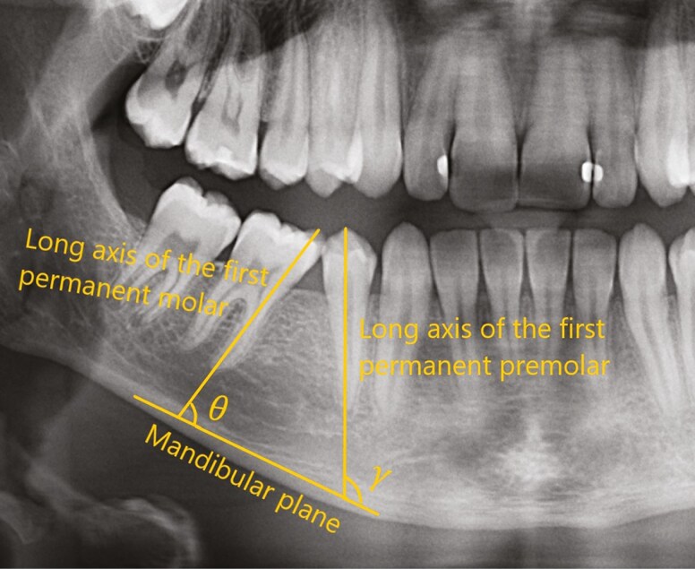
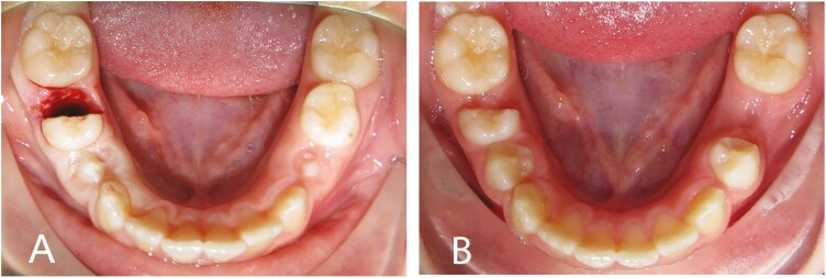
Materials and methods: Patients with bilateral agenesis of the second mandibular molars and unerupted second molars were included and randomly allocated to either extraction or hemisection on the left or right side of the mandible. Clinical and radiographic examinations were conducted at baseline (T1) and after a mean follow-up period of 4.2 years (T2). Measurements of the residual spaces and tooth angulation of the mandibular first molar and premolar following extraction were blinded assessed on panoramic radiographs and cast models. The number of visits, chair time, social costs, and direct and indirect costs were calculated using cost minimisation analysis.
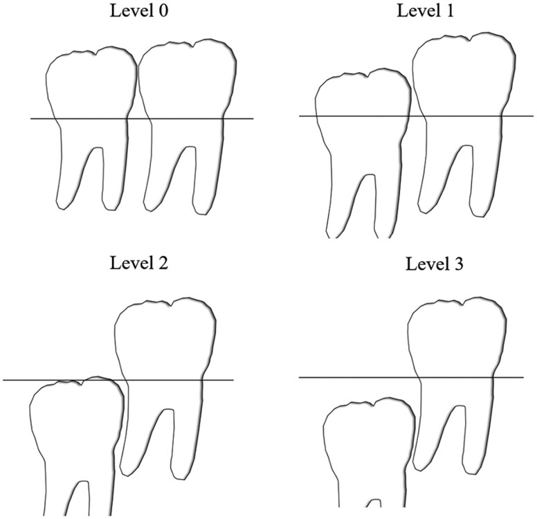
Results: A total of 40 patients (25 boys and 15 girls) with a mean age of 10.03 ± 1.07 years at T1 participated. No patient was lost to follow-up. The residual space between the first permanent molar and the first permanent premolar was 2.04 ± 1.67 mm for hemisection and 2.39 ± 1.86 mm for extraction (p = 0.053). A larger residual space was observed between the first permanent premolar and the canine on the hemisection side (1.80 ± 1.01 mm) than on the extraction side (1.55 ± 0.92 mm), (p = 0.045). No difference was found between the interventions regarding the angulation of the first permanent molar (p = 0.0914) or the angulation of the first permanent premolar (p = 0.7812). Hemisection resulted in significantly more complications (p = 0.0176) and was associated with substantially higher material costs, more chair time and higher indirect costs than conventional extraction (p < 0.0001).
Conclusion: Hemisection is not recommended as an interceptive extraction option for patients with congenitally missing mandibular second premolars, as only minimal, clinically irrelevant differences were observed compared with conventional extraction. Moreover, hemisection is associated with increased costs and a higher risk of complications.
Trial registration: The trial was registered with https://www.researchweb.org/is/sverige, registration number: 967125.
期刊介绍:
The European Journal of Orthodontics publishes papers of excellence on all aspects of orthodontics including craniofacial development and growth. The emphasis of the journal is on full research papers. Succinct and carefully prepared papers are favoured in terms of impact as well as readability.
 求助内容:
求助内容: 应助结果提醒方式:


