{"title":"Influence of L-arginine on hydroxyapatite-based ovine bone graft - An <i>in vitro</i> evaluation of surface characteristics and cell viability.","authors":"Maaz Vohra, Subhabrata Maiti, Khushali K Shah, Lokitha Raju, Deepak Nallaswamy, Rajalakshmanan Eswaramoorthy","doi":"10.4103/drj.drj_263_24","DOIUrl":null,"url":null,"abstract":"<p><strong>Background: </strong>Current challenges in bone grafting revolve around the limited availability of autografts and the complications associated with their use. Promising alternatives include osteoinductive substances stimulating stem cells to mature into bone-forming osteoblasts. However, existing products lack optimal characteristics of a bone graft. The study aimed to evaluate the impact of L-arginine treatment on hydroxyapatite (HA) derived from ovine bone and compare its surface and mechanical properties to that of the commercially available xenograft-Bio-Oss.</p><p><strong>Materials and methods: </strong>The research was structured as an <i>in vitro</i> investigation, wherein HA was formulated from ovine bone. The sintering process involved heating it to 360°C for 3 h and adding the amino acid L-arginine. Different tests were done which included scanning electron microscopy (SEM), X-ray diffractometry, Fourier-transform infrared spectroscopy (FTIR), and 3-(4,5-dimethylthiazol-2-yl)-2,5-diphenyltetrazolium bromide assay. The goal was to compare these results with a commercially available bone graft called BioOss, especially regarding their physical and chemical characteristics. Data were analyzed in SPSS software using one way ANOVA test, significant level at 0.05.</p><p><strong>Results: </strong>A bone graft made of HA and L-arginine displayed a complex and interconnected pore structure, indicating that the sintering process had a significant impact. SEM confirmed this. FTIR analysis identified peaks at 650-700 cm<sup>-1</sup> and 1000-1100 cm<sup>-1</sup>, confirming the presence of HA and L-arginine. X-ray Diffraction assessments also confirmed the existence of both substances in the sintered specimens, supporting their suitability for various biomedical applications.</p><p><strong>Conclusion: </strong>The study presents a novel approach, deproteinizing a bone graft followed by sintering at 360°C with L-arginine. Physicochemical analyses confirmed desired mechanical attributes and surface characteristics. Further investigations are needed to evaluate cellular adherence, immunological response, and osteogenesis in relevant animal models.</p>","PeriodicalId":11016,"journal":{"name":"Dental Research Journal","volume":"22 ","pages":"19"},"PeriodicalIF":0.0000,"publicationDate":"2025-05-22","publicationTypes":"Journal Article","fieldsOfStudy":null,"isOpenAccess":false,"openAccessPdf":"https://www.ncbi.nlm.nih.gov/pmc/articles/PMC12155389/pdf/","citationCount":"0","resultStr":null,"platform":"Semanticscholar","paperid":null,"PeriodicalName":"Dental Research Journal","FirstCategoryId":"1085","ListUrlMain":"https://doi.org/10.4103/drj.drj_263_24","RegionNum":0,"RegionCategory":null,"ArticlePicture":[],"TitleCN":null,"AbstractTextCN":null,"PMCID":null,"EPubDate":"2025/1/1 0:00:00","PubModel":"eCollection","JCR":"Q2","JCRName":"Dentistry","Score":null,"Total":0}
引用次数: 0
Abstract
Background: Current challenges in bone grafting revolve around the limited availability of autografts and the complications associated with their use. Promising alternatives include osteoinductive substances stimulating stem cells to mature into bone-forming osteoblasts. However, existing products lack optimal characteristics of a bone graft. The study aimed to evaluate the impact of L-arginine treatment on hydroxyapatite (HA) derived from ovine bone and compare its surface and mechanical properties to that of the commercially available xenograft-Bio-Oss.
Materials and methods: The research was structured as an in vitro investigation, wherein HA was formulated from ovine bone. The sintering process involved heating it to 360°C for 3 h and adding the amino acid L-arginine. Different tests were done which included scanning electron microscopy (SEM), X-ray diffractometry, Fourier-transform infrared spectroscopy (FTIR), and 3-(4,5-dimethylthiazol-2-yl)-2,5-diphenyltetrazolium bromide assay. The goal was to compare these results with a commercially available bone graft called BioOss, especially regarding their physical and chemical characteristics. Data were analyzed in SPSS software using one way ANOVA test, significant level at 0.05.
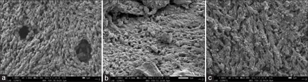
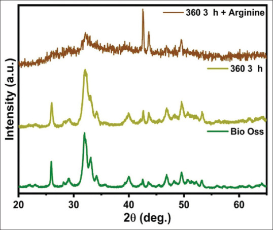
Results: A bone graft made of HA and L-arginine displayed a complex and interconnected pore structure, indicating that the sintering process had a significant impact. SEM confirmed this. FTIR analysis identified peaks at 650-700 cm-1 and 1000-1100 cm-1, confirming the presence of HA and L-arginine. X-ray Diffraction assessments also confirmed the existence of both substances in the sintered specimens, supporting their suitability for various biomedical applications.
Conclusion: The study presents a novel approach, deproteinizing a bone graft followed by sintering at 360°C with L-arginine. Physicochemical analyses confirmed desired mechanical attributes and surface characteristics. Further investigations are needed to evaluate cellular adherence, immunological response, and osteogenesis in relevant animal models.
期刊介绍:
Dental Research Journal, a publication of Isfahan University of Medical Sciences, is a peer-reviewed online journal with Bimonthly print on demand compilation of issues published. The journal’s full text is available online at http://www.drjjournal.net. The journal allows free access (Open Access) to its contents and permits authors to self-archive final accepted version of the articles on any OAI-compliant institutional / subject-based repository. The journal will cover technical and clinical studies related to health, ethical and social issues in field of Dentistry. Articles with clinical interest and implications will be given preference.


 求助内容:
求助内容: 应助结果提醒方式:


