The Biofunctional Monomer, the Calcium Salt of 4-Methacryloxyethyl Trimellitic Acid, Promotes Odontoblast Differentiation in Three-Dimensional Culture System.
{"title":"The Biofunctional Monomer, the Calcium Salt of 4-Methacryloxyethyl Trimellitic Acid, Promotes Odontoblast Differentiation in Three-Dimensional Culture System.","authors":"Yaxin Rao, Youjing Qiu, Takashi Saito","doi":"10.1155/ijbm/3693662","DOIUrl":null,"url":null,"abstract":"<p><p>This study evaluated the effects of the biofunctional monomer CMET on the proliferation, differentiation, and mineralization of MDPC-23, odontoblast-like cells in a three-dimensional (3D) culture system using type I collagen. CMET (0.3%, w/v) facilitated the early adhesion and spreading of the cells in type I collagen gels. It significantly promoted cell proliferation in 0.2% and 0.3% concentrations. ALP activity also increased in the 0.3% CMET group. The 0.3% CMET group markedly enhanced odontogenic differentiation by upregulating mRNA of odontogenic differentiation markers such as DSPP and DSP-1. Mineral nodule formation in MDPC-23 cells grown in the 0.3% CMET group was markedly increased compared to that in the control group. After treating the cells with the three MAPK inhibitors, the ability of CMET to stimulate ALP activity in MDPC-23 cells was totally suppressed to control levels by the p38 inhibitor, SB202190. The enhancement of mineralization of MDPC-23 by CMET was partially impeded by SB202190. The results demonstrated that the biofunctional monomer CMET induced proliferation, differentiation, and mineralization of odontoblast-like cells in a 3D culture system using type I collagen gel at a concentration of 0.3%. Thus, combining CMET and type I collagen gel as a scaffold does not exhibit apparent cytotoxicity and is suggested to have immense potential for dentin regeneration.</p>","PeriodicalId":13704,"journal":{"name":"International Journal of Biomaterials","volume":"2025 ","pages":"3693662"},"PeriodicalIF":4.5000,"publicationDate":"2025-06-03","publicationTypes":"Journal Article","fieldsOfStudy":null,"isOpenAccess":false,"openAccessPdf":"https://www.ncbi.nlm.nih.gov/pmc/articles/PMC12151624/pdf/","citationCount":"0","resultStr":null,"platform":"Semanticscholar","paperid":null,"PeriodicalName":"International Journal of Biomaterials","FirstCategoryId":"1085","ListUrlMain":"https://doi.org/10.1155/ijbm/3693662","RegionNum":0,"RegionCategory":null,"ArticlePicture":[],"TitleCN":null,"AbstractTextCN":null,"PMCID":null,"EPubDate":"2025/1/1 0:00:00","PubModel":"eCollection","JCR":"Q3","JCRName":"MATERIALS SCIENCE, BIOMATERIALS","Score":null,"Total":0}
引用次数: 0
Abstract
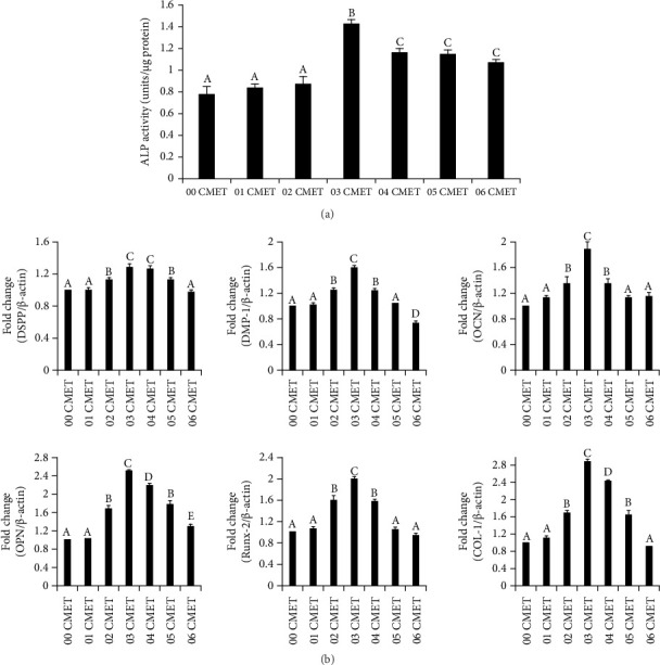
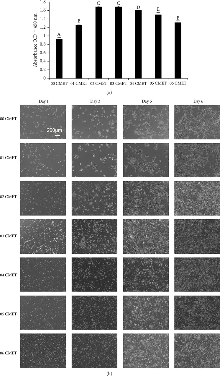
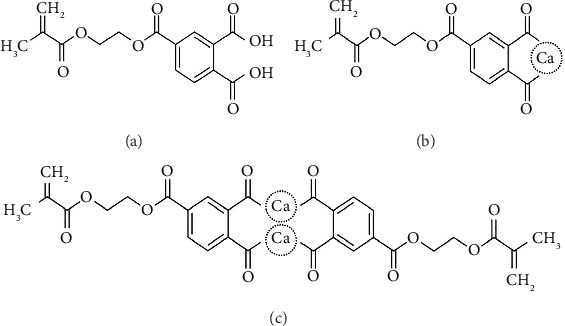
This study evaluated the effects of the biofunctional monomer CMET on the proliferation, differentiation, and mineralization of MDPC-23, odontoblast-like cells in a three-dimensional (3D) culture system using type I collagen. CMET (0.3%, w/v) facilitated the early adhesion and spreading of the cells in type I collagen gels. It significantly promoted cell proliferation in 0.2% and 0.3% concentrations. ALP activity also increased in the 0.3% CMET group. The 0.3% CMET group markedly enhanced odontogenic differentiation by upregulating mRNA of odontogenic differentiation markers such as DSPP and DSP-1. Mineral nodule formation in MDPC-23 cells grown in the 0.3% CMET group was markedly increased compared to that in the control group. After treating the cells with the three MAPK inhibitors, the ability of CMET to stimulate ALP activity in MDPC-23 cells was totally suppressed to control levels by the p38 inhibitor, SB202190. The enhancement of mineralization of MDPC-23 by CMET was partially impeded by SB202190. The results demonstrated that the biofunctional monomer CMET induced proliferation, differentiation, and mineralization of odontoblast-like cells in a 3D culture system using type I collagen gel at a concentration of 0.3%. Thus, combining CMET and type I collagen gel as a scaffold does not exhibit apparent cytotoxicity and is suggested to have immense potential for dentin regeneration.

 求助内容:
求助内容: 应助结果提醒方式:


