David J Williamson, Cecilia Zaza, Irene Carlon-Andres, Tobias Starling, Alessia Gentili, Joseph W Thrush, Audrey Le Bas, Ravi Teja Ravi, Stuart Neil, Ray J Owens, Maud Dumoux, Sabrina Simoncelli, Sergi Padilla-Parra
{"title":"Single-molecule localisation microscopy approaches reveal envelope glycoprotein clusters in single-enveloped viruses: a potential functional role?","authors":"David J Williamson, Cecilia Zaza, Irene Carlon-Andres, Tobias Starling, Alessia Gentili, Joseph W Thrush, Audrey Le Bas, Ravi Teja Ravi, Stuart Neil, Ray J Owens, Maud Dumoux, Sabrina Simoncelli, Sergi Padilla-Parra","doi":"10.1042/BST20240769","DOIUrl":null,"url":null,"abstract":"<p><p>Understanding how viruses enter and fuse with host cells is crucial for developing effective antiviral therapies. The process of viral entry and fusion involves a series of complex steps that allow the virus to breach the host cell membrane and deliver its genetic material inside, with viral fusogens often co-operating to attain the required energy for successful membrane fusion. This co-operative clustering of fusogens in viral envelopes is similar to receptor clustering in cellular systems, where receptors aggregate to initiate signalling cascades. Single-molecule localisation microscopy (SMLM) approaches have emerged as powerful tools to study these intricate mechanisms, allowing the observation of proteins with unprecedented levels of detail. These technologies provide unparalleled insights into the dynamics of viral entry and fusion at a molecular level, revealing how the co-ordinated action of fusogens facilitates membrane fusion. By employing the newest advances in SMLM techniques, such as DNA-PAINT and MINFLUX, we anticipate that precise information on the key steps of viral fusion can be revealed with high spatial and temporal resolutions, identifying critical points in the process that can be targeted by antiviral strategies.</p>","PeriodicalId":8841,"journal":{"name":"Biochemical Society transactions","volume":" ","pages":"643-652"},"PeriodicalIF":4.3000,"publicationDate":"2025-06-30","publicationTypes":"Journal Article","fieldsOfStudy":null,"isOpenAccess":false,"openAccessPdf":"https://www.ncbi.nlm.nih.gov/pmc/articles/PMC12236106/pdf/","citationCount":"0","resultStr":null,"platform":"Semanticscholar","paperid":null,"PeriodicalName":"Biochemical Society transactions","FirstCategoryId":"99","ListUrlMain":"https://doi.org/10.1042/BST20240769","RegionNum":3,"RegionCategory":"生物学","ArticlePicture":[],"TitleCN":null,"AbstractTextCN":null,"PMCID":null,"EPubDate":"","PubModel":"","JCR":"Q2","JCRName":"BIOCHEMISTRY & MOLECULAR BIOLOGY","Score":null,"Total":0}
引用次数: 0
Abstract
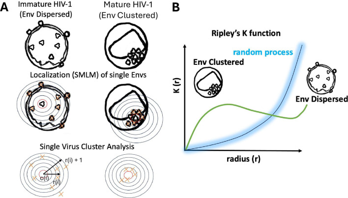
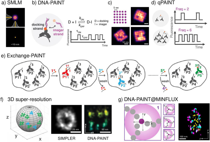
Understanding how viruses enter and fuse with host cells is crucial for developing effective antiviral therapies. The process of viral entry and fusion involves a series of complex steps that allow the virus to breach the host cell membrane and deliver its genetic material inside, with viral fusogens often co-operating to attain the required energy for successful membrane fusion. This co-operative clustering of fusogens in viral envelopes is similar to receptor clustering in cellular systems, where receptors aggregate to initiate signalling cascades. Single-molecule localisation microscopy (SMLM) approaches have emerged as powerful tools to study these intricate mechanisms, allowing the observation of proteins with unprecedented levels of detail. These technologies provide unparalleled insights into the dynamics of viral entry and fusion at a molecular level, revealing how the co-ordinated action of fusogens facilitates membrane fusion. By employing the newest advances in SMLM techniques, such as DNA-PAINT and MINFLUX, we anticipate that precise information on the key steps of viral fusion can be revealed with high spatial and temporal resolutions, identifying critical points in the process that can be targeted by antiviral strategies.
期刊介绍:
Biochemical Society Transactions is the reviews journal of the Biochemical Society. Publishing concise reviews written by experts in the field, providing a timely snapshot of the latest developments across all areas of the molecular and cellular biosciences.
Elevating our authors’ ideas and expertise, each review includes a perspectives section where authors offer comment on the latest advances, a glimpse of future challenges and highlighting the importance of associated research areas in far broader contexts.
 求助内容:
求助内容: 应助结果提醒方式:


