{"title":"Advancing extracellular vesicle production: improving physiological relevance and yield with 3D cell culture","authors":"Kara Cook and Huiyan Li","doi":"10.1039/D5NR00707K","DOIUrl":null,"url":null,"abstract":"<p >Extracellular vesicles (EVs) are essential nanoscale mediators of intercellular communication, holding significant potential as early disease biomarkers and therapeutic agents. Present in biological fluids like blood, EVs and their molecular cargo can be detected in liquid biopsies for diverse diagnostic and therapeutic applications. However, the availability of patient samples is often limited for such research. To tackle this challenge and gain insights into <em>in vivo</em> disease mechanisms, <em>in vitro</em> production of EVs from the cell culture models that closely mimic <em>in vivo</em> conditions has become an essential tool. While 2D cell culture has been the standard for high-throughput studies for decades, 3D cell culture is emerging as a more physiologically relevant <em>in vitro</em> tool for mimicking <em>in vivo</em> environments and providing deeper insights into disease. However, there is currently a lack of literature synthesizing and comparing the effects of 3D <em>versus</em> 2D cell culture models on EV production and analysis. In this review, we examine recent studies that compare the impacts of 3D and 2D cell culture models on EV yield, composition, and functionality. We categorize 3D models into subtypes, including spheroids, hydrogels, rigid scaffolds, and bioreactors. Details of each model's impact on EVs compared to 2D cell culture are presented. Furthermore, we discuss the advantages and limitations of these 3D models as identified in individual studies, offering insights to guide future research directions in this evolving field.</p>","PeriodicalId":92,"journal":{"name":"Nanoscale","volume":" 25","pages":" 15110-15131"},"PeriodicalIF":5.1000,"publicationDate":"2025-06-10","publicationTypes":"Journal Article","fieldsOfStudy":null,"isOpenAccess":false,"openAccessPdf":"https://pubs.rsc.org/en/content/articlepdf/2025/nr/d5nr00707k?page=search","citationCount":"0","resultStr":null,"platform":"Semanticscholar","paperid":null,"PeriodicalName":"Nanoscale","FirstCategoryId":"88","ListUrlMain":"https://pubs.rsc.org/en/content/articlelanding/2025/nr/d5nr00707k","RegionNum":3,"RegionCategory":"材料科学","ArticlePicture":[],"TitleCN":null,"AbstractTextCN":null,"PMCID":null,"EPubDate":"","PubModel":"","JCR":"Q1","JCRName":"CHEMISTRY, MULTIDISCIPLINARY","Score":null,"Total":0}
引用次数: 0
Abstract
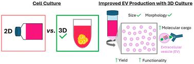
Extracellular vesicles (EVs) are essential nanoscale mediators of intercellular communication, holding significant potential as early disease biomarkers and therapeutic agents. Present in biological fluids like blood, EVs and their molecular cargo can be detected in liquid biopsies for diverse diagnostic and therapeutic applications. However, the availability of patient samples is often limited for such research. To tackle this challenge and gain insights into in vivo disease mechanisms, in vitro production of EVs from the cell culture models that closely mimic in vivo conditions has become an essential tool. While 2D cell culture has been the standard for high-throughput studies for decades, 3D cell culture is emerging as a more physiologically relevant in vitro tool for mimicking in vivo environments and providing deeper insights into disease. However, there is currently a lack of literature synthesizing and comparing the effects of 3D versus 2D cell culture models on EV production and analysis. In this review, we examine recent studies that compare the impacts of 3D and 2D cell culture models on EV yield, composition, and functionality. We categorize 3D models into subtypes, including spheroids, hydrogels, rigid scaffolds, and bioreactors. Details of each model's impact on EVs compared to 2D cell culture are presented. Furthermore, we discuss the advantages and limitations of these 3D models as identified in individual studies, offering insights to guide future research directions in this evolving field.
期刊介绍:
Nanoscale is a high-impact international journal, publishing high-quality research across nanoscience and nanotechnology. Nanoscale publishes a full mix of research articles on experimental and theoretical work, including reviews, communications, and full papers.Highly interdisciplinary, this journal appeals to scientists, researchers and professionals interested in nanoscience and nanotechnology, quantum materials and quantum technology, including the areas of physics, chemistry, biology, medicine, materials, energy/environment, information technology, detection science, healthcare and drug discovery, and electronics.
 求助内容:
求助内容: 应助结果提醒方式:


