Erin P Snoddy, Tien T Tang, Natalie Fowlkes, Thomas Huynh, Kari J Brewer Savannah, Alejandro Contreras, Gregory Reece, Kristy K Brock
{"title":"Development of an H&E on-block staining technique for collagen detection in cryo-fluorescence tomography imaging of frozen breast tissue samples.","authors":"Erin P Snoddy, Tien T Tang, Natalie Fowlkes, Thomas Huynh, Kari J Brewer Savannah, Alejandro Contreras, Gregory Reece, Kristy K Brock","doi":"10.1371/journal.pone.0324493","DOIUrl":null,"url":null,"abstract":"<p><p>Hematoxylin and eosin (H&E) staining is widely considered to be the gold-standard diagnostic tool for histopathology evaluation. However, the fatty nature of some tissue types, such as breast tissue, presents challenges with cryo-sectioning, often resulting in artifacts that can make histopathologic interpretation and correlation with other imaging modalities virtually impossible. We present an optimized on-block H&E staining technique that improves contrast for identifying collagenous stroma during cryo-fluorescence tomography (CFT) sectioning. In this prospective study, we embedded four breast specimens with confirmed ligaments from a bilateral mastopexy in an optimal cutting temperature block. Two of the samples were processed on a CFT imager and stained with our on-block staining protocol. In this protocol, hematoxylin was applied to the block-face before being washed with deionized water. Eosin was then applied and washed with 95% ethanol. We then applied mounting medium and acquired images with a stereo-dissecting microscope and camera. Prior to staining, GFP fluorescence and white-light images were acquired with the CFT system to serve as a validation metric. The other two samples were sectioned on a standard cryostat and stained according to gold-standard H&E protocol. The resulting microscope slides were imaged with a digital slide scanner and viewed with Leica Imagescope software. An experienced pathologist evaluated both sets of images for qualitative comparisons. Pathologist evaluation confirmed that striations from on-block staining were qualitatively comparable with collagen tracks identified in gold-standard histology images. Furthermore, GFP images captured collagen autofluorescence, which aligned with the same structures identified by our on-block staining protocol. Our on-block staining technique shows comparable visualization of collagenous structures at the mesoscopic level for fresh breast tissue samples. This technique improves tissue contrast and region of interest selection for histology during CFT imaging for analysis of the stromal architecture of the breast.</p>","PeriodicalId":20189,"journal":{"name":"PLoS ONE","volume":"20 6","pages":"e0324493"},"PeriodicalIF":2.6000,"publicationDate":"2025-06-09","publicationTypes":"Journal Article","fieldsOfStudy":null,"isOpenAccess":false,"openAccessPdf":"https://www.ncbi.nlm.nih.gov/pmc/articles/PMC12148110/pdf/","citationCount":"0","resultStr":null,"platform":"Semanticscholar","paperid":null,"PeriodicalName":"PLoS ONE","FirstCategoryId":"103","ListUrlMain":"https://doi.org/10.1371/journal.pone.0324493","RegionNum":3,"RegionCategory":"综合性期刊","ArticlePicture":[],"TitleCN":null,"AbstractTextCN":null,"PMCID":null,"EPubDate":"2025/1/1 0:00:00","PubModel":"eCollection","JCR":"Q1","JCRName":"MULTIDISCIPLINARY SCIENCES","Score":null,"Total":0}
引用次数: 0
Abstract
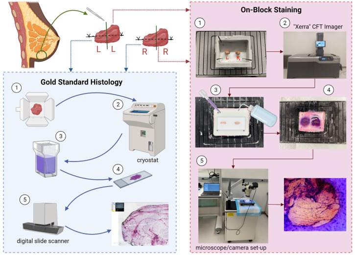
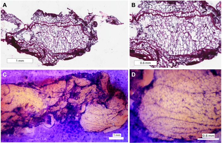
Hematoxylin and eosin (H&E) staining is widely considered to be the gold-standard diagnostic tool for histopathology evaluation. However, the fatty nature of some tissue types, such as breast tissue, presents challenges with cryo-sectioning, often resulting in artifacts that can make histopathologic interpretation and correlation with other imaging modalities virtually impossible. We present an optimized on-block H&E staining technique that improves contrast for identifying collagenous stroma during cryo-fluorescence tomography (CFT) sectioning. In this prospective study, we embedded four breast specimens with confirmed ligaments from a bilateral mastopexy in an optimal cutting temperature block. Two of the samples were processed on a CFT imager and stained with our on-block staining protocol. In this protocol, hematoxylin was applied to the block-face before being washed with deionized water. Eosin was then applied and washed with 95% ethanol. We then applied mounting medium and acquired images with a stereo-dissecting microscope and camera. Prior to staining, GFP fluorescence and white-light images were acquired with the CFT system to serve as a validation metric. The other two samples were sectioned on a standard cryostat and stained according to gold-standard H&E protocol. The resulting microscope slides were imaged with a digital slide scanner and viewed with Leica Imagescope software. An experienced pathologist evaluated both sets of images for qualitative comparisons. Pathologist evaluation confirmed that striations from on-block staining were qualitatively comparable with collagen tracks identified in gold-standard histology images. Furthermore, GFP images captured collagen autofluorescence, which aligned with the same structures identified by our on-block staining protocol. Our on-block staining technique shows comparable visualization of collagenous structures at the mesoscopic level for fresh breast tissue samples. This technique improves tissue contrast and region of interest selection for histology during CFT imaging for analysis of the stromal architecture of the breast.
期刊介绍:
PLOS ONE is an international, peer-reviewed, open-access, online publication. PLOS ONE welcomes reports on primary research from any scientific discipline. It provides:
* Open-access—freely accessible online, authors retain copyright
* Fast publication times
* Peer review by expert, practicing researchers
* Post-publication tools to indicate quality and impact
* Community-based dialogue on articles
* Worldwide media coverage

 求助内容:
求助内容: 应助结果提醒方式:


