Comparison of lesion segmentation performance in diffusion-weighted imaging and apparent diffusion coefficient images of stroke by artificial neural networks.
Seok Jin Bang, Yong-Tae Kim, Young Jae Kim, Kwang Gi Kim
{"title":"Comparison of lesion segmentation performance in diffusion-weighted imaging and apparent diffusion coefficient images of stroke by artificial neural networks.","authors":"Seok Jin Bang, Yong-Tae Kim, Young Jae Kim, Kwang Gi Kim","doi":"10.1371/journal.pone.0324021","DOIUrl":null,"url":null,"abstract":"<p><p>Stroke is the second leading cause of death, accounting for 11% of deaths worldwide. Comparing diffusion-weighted imaging (DWI) and apparent diffusion coefficient (ADC) images is important for stroke diagnosis, but most studies have focused on lesion segmentation using DWI. In this study, we compared the performance of lesion segmentation using DWI and ADC images. This study was conducted using a retrospective design A dataset was constructed using data from 360 patients with ischemic stroke collected from Gachon University Gil Medical Center. Artificial intelligence models, U-Net, and a fully connected network (FCN), were used to train each type of image data. The performance of the models was validated using five-fold cross-validation and evaluated based on metrics such as the dice similarity coefficient (DSC), accuracy, precision, and recall. As a result, the U-Net model demonstrated a DSC of 92.13 ± 0.91% on DWI and 83.68 ± 10% on ADC, whereas the FCN model exhibited a DSC of 82.86 ± 1.56% on DWI and 79.26 ± 1.19% on ADC. These metrics indicated that the trained models were suitable for lesion segmentation. A comparative analysis of DWI and ADC based on the trained models revealed similar results across the models, suggesting that lesion segmentation on ADC images is appropriate. For future research, the accuracy of ADC images is recommended to be imporved by utilizing images with different b-values, or training models with datasets that combe DWI and ADC images based on enhanced data.</p>","PeriodicalId":20189,"journal":{"name":"PLoS ONE","volume":"20 6","pages":"e0324021"},"PeriodicalIF":2.6000,"publicationDate":"2025-06-09","publicationTypes":"Journal Article","fieldsOfStudy":null,"isOpenAccess":false,"openAccessPdf":"https://www.ncbi.nlm.nih.gov/pmc/articles/PMC12148192/pdf/","citationCount":"0","resultStr":null,"platform":"Semanticscholar","paperid":null,"PeriodicalName":"PLoS ONE","FirstCategoryId":"103","ListUrlMain":"https://doi.org/10.1371/journal.pone.0324021","RegionNum":3,"RegionCategory":"综合性期刊","ArticlePicture":[],"TitleCN":null,"AbstractTextCN":null,"PMCID":null,"EPubDate":"2025/1/1 0:00:00","PubModel":"eCollection","JCR":"Q1","JCRName":"MULTIDISCIPLINARY SCIENCES","Score":null,"Total":0}
引用次数: 0
Abstract
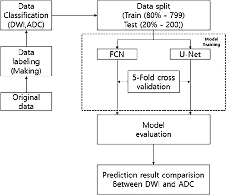
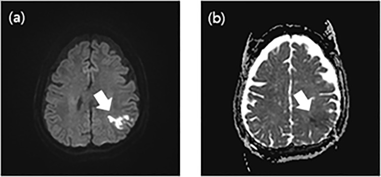
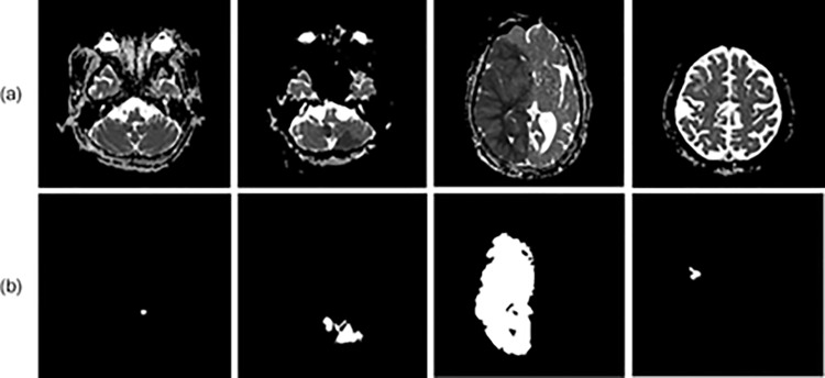
Stroke is the second leading cause of death, accounting for 11% of deaths worldwide. Comparing diffusion-weighted imaging (DWI) and apparent diffusion coefficient (ADC) images is important for stroke diagnosis, but most studies have focused on lesion segmentation using DWI. In this study, we compared the performance of lesion segmentation using DWI and ADC images. This study was conducted using a retrospective design A dataset was constructed using data from 360 patients with ischemic stroke collected from Gachon University Gil Medical Center. Artificial intelligence models, U-Net, and a fully connected network (FCN), were used to train each type of image data. The performance of the models was validated using five-fold cross-validation and evaluated based on metrics such as the dice similarity coefficient (DSC), accuracy, precision, and recall. As a result, the U-Net model demonstrated a DSC of 92.13 ± 0.91% on DWI and 83.68 ± 10% on ADC, whereas the FCN model exhibited a DSC of 82.86 ± 1.56% on DWI and 79.26 ± 1.19% on ADC. These metrics indicated that the trained models were suitable for lesion segmentation. A comparative analysis of DWI and ADC based on the trained models revealed similar results across the models, suggesting that lesion segmentation on ADC images is appropriate. For future research, the accuracy of ADC images is recommended to be imporved by utilizing images with different b-values, or training models with datasets that combe DWI and ADC images based on enhanced data.
期刊介绍:
PLOS ONE is an international, peer-reviewed, open-access, online publication. PLOS ONE welcomes reports on primary research from any scientific discipline. It provides:
* Open-access—freely accessible online, authors retain copyright
* Fast publication times
* Peer review by expert, practicing researchers
* Post-publication tools to indicate quality and impact
* Community-based dialogue on articles
* Worldwide media coverage

 求助内容:
求助内容: 应助结果提醒方式:


