Hellen Carvalho Ribeiro, William Paganini Mayer, Jacob Matz, Josemberg da Silva Baptista
{"title":"Posterior Malleolus: Morphologic Classification, Morphometry, and Clinical Insights.","authors":"Hellen Carvalho Ribeiro, William Paganini Mayer, Jacob Matz, Josemberg da Silva Baptista","doi":"10.1177/24730114251341900","DOIUrl":null,"url":null,"abstract":"<p><strong>Background: </strong>In this study, we provide a comprehensive description of the morphometrics of the distal tibiae and propose that the intact posterior malleolus (PM) exhibits clinically relevant morphologic variation. These differences may have implications for fracture classification, fixation strategy, and implant design.</p><p><strong>Methods: </strong>Fifty-two isolated dry tibias were analyzed to determine the PM morphometric parameters. Five key morphometric points were identified, and the PM was defined as the posterior bony projection of the distal tibial epiphysis. The malleolar groove established the PM's medial limitation, the posterior portion of the fibular notch defined the lateral limit, and the anterior boundary was a line connecting these landmarks across the inferior articular surface. PM shapes were categorized based on consistent morphologic patterns. Cross-sections of the distal tibia were performed to assess trabecular bone morphology and density.</p><p><strong>Results: </strong>We found the PM presenting 3 distinct morphologic types: rounded, triangular, and trapezoid. Triangular and trapezoid types exhibited larger dimensions and robust bone tissue, whereas tibias with a rounded PM displayed smaller dimensions and delicate bone architecture.</p><p><strong>Conclusion: </strong>These novel findings reveal PM morphologic diversity, which may enhance our understanding of PM fracture patterns and optimize the development of surgical implants.</p>","PeriodicalId":12429,"journal":{"name":"Foot & Ankle Orthopaedics","volume":"10 2","pages":"24730114251341900"},"PeriodicalIF":0.0000,"publicationDate":"2025-06-08","publicationTypes":"Journal Article","fieldsOfStudy":null,"isOpenAccess":false,"openAccessPdf":"https://www.ncbi.nlm.nih.gov/pmc/articles/PMC12146590/pdf/","citationCount":"0","resultStr":null,"platform":"Semanticscholar","paperid":null,"PeriodicalName":"Foot & Ankle Orthopaedics","FirstCategoryId":"1085","ListUrlMain":"https://doi.org/10.1177/24730114251341900","RegionNum":0,"RegionCategory":null,"ArticlePicture":[],"TitleCN":null,"AbstractTextCN":null,"PMCID":null,"EPubDate":"2025/4/1 0:00:00","PubModel":"eCollection","JCR":"","JCRName":"","Score":null,"Total":0}
引用次数: 0
Abstract
Background: In this study, we provide a comprehensive description of the morphometrics of the distal tibiae and propose that the intact posterior malleolus (PM) exhibits clinically relevant morphologic variation. These differences may have implications for fracture classification, fixation strategy, and implant design.
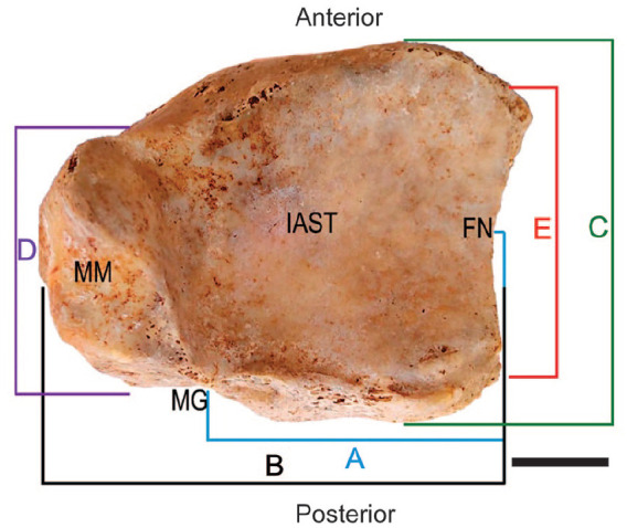
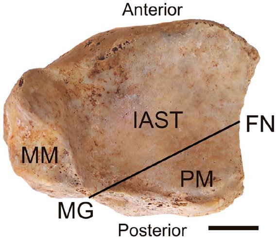
Methods: Fifty-two isolated dry tibias were analyzed to determine the PM morphometric parameters. Five key morphometric points were identified, and the PM was defined as the posterior bony projection of the distal tibial epiphysis. The malleolar groove established the PM's medial limitation, the posterior portion of the fibular notch defined the lateral limit, and the anterior boundary was a line connecting these landmarks across the inferior articular surface. PM shapes were categorized based on consistent morphologic patterns. Cross-sections of the distal tibia were performed to assess trabecular bone morphology and density.
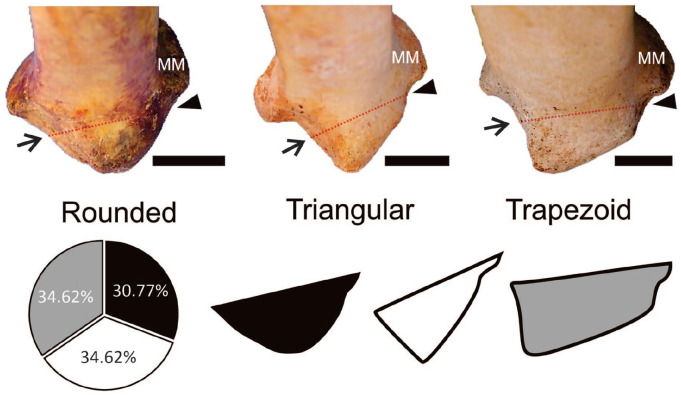
Results: We found the PM presenting 3 distinct morphologic types: rounded, triangular, and trapezoid. Triangular and trapezoid types exhibited larger dimensions and robust bone tissue, whereas tibias with a rounded PM displayed smaller dimensions and delicate bone architecture.
Conclusion: These novel findings reveal PM morphologic diversity, which may enhance our understanding of PM fracture patterns and optimize the development of surgical implants.

 求助内容:
求助内容: 应助结果提醒方式:


