{"title":"Contribution of 3D visualization and printing in teaching lung segments anatomy.","authors":"Gabrielle Drevet, Valentin Soldea, Sylvain Gouttard, Melia Virely, Jean-Michel Maury, François Tronc","doi":"10.1186/s41205-025-00272-z","DOIUrl":null,"url":null,"abstract":"<p><strong>Background: </strong>The knowledge and understanding of the anatomy of lung segments is of great importance while segmentectomies are increasingly performed. To introduce new technologies and tools in anatomy teaching could help students to improve their skills.</p><p><strong>Methods: </strong>Students participants (n = 16) were divided into 3 groups: traditional (n = 5), 3D visualization (n = 5) and 3D printing group (n = 6). Each student took a pre- and post-test exam. The traditional teaching group had lessons using 2D anatomical drawings, the 3D visualization group had lessons using a dedicated software allowing anatomical 3D reconstructions and the 3D printing group had lessons using 3D printed anatomical models.</p><p><strong>Results: </strong>Students of the whole cohort had significant better scores at the post test (mean score = 14.2) compared to the pretest (mean score = 7.9) (p = 0.0011). In the traditional and 3D printing groups, students had significant better scores in the post-test (mean scores = 17.7 and 14.2 respectively) than in the pre-test (mean scores = 8.2 and 7.5; p = 0.0247 and p = 0.0003 respectively). There was no significant difference between the pre and post-test scores for the 3D visualization group (mean score = 8.2 and 11.7 respectively) (p = 0.4347).</p><p><strong>Conclusions: </strong>The knowledge of lung segment anatomy is poor among our medical students. Both traditional and 3D-printed teaching was shown effective. The contribution of 3D printed models would probably improve anatomy teaching among medical students. The introduction of this technology is instinctive and easy to use for both students and teachers. Furthermore, this technique was not particularly expensive to set up.</p>","PeriodicalId":72036,"journal":{"name":"3D printing in medicine","volume":"11 1","pages":"25"},"PeriodicalIF":3.1000,"publicationDate":"2025-06-09","publicationTypes":"Journal Article","fieldsOfStudy":null,"isOpenAccess":false,"openAccessPdf":"https://www.ncbi.nlm.nih.gov/pmc/articles/PMC12147242/pdf/","citationCount":"0","resultStr":null,"platform":"Semanticscholar","paperid":null,"PeriodicalName":"3D printing in medicine","FirstCategoryId":"1085","ListUrlMain":"https://doi.org/10.1186/s41205-025-00272-z","RegionNum":0,"RegionCategory":null,"ArticlePicture":[],"TitleCN":null,"AbstractTextCN":null,"PMCID":null,"EPubDate":"","PubModel":"","JCR":"Q1","JCRName":"RADIOLOGY, NUCLEAR MEDICINE & MEDICAL IMAGING","Score":null,"Total":0}
引用次数: 0
Abstract
Background: The knowledge and understanding of the anatomy of lung segments is of great importance while segmentectomies are increasingly performed. To introduce new technologies and tools in anatomy teaching could help students to improve their skills.
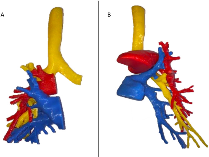
Methods: Students participants (n = 16) were divided into 3 groups: traditional (n = 5), 3D visualization (n = 5) and 3D printing group (n = 6). Each student took a pre- and post-test exam. The traditional teaching group had lessons using 2D anatomical drawings, the 3D visualization group had lessons using a dedicated software allowing anatomical 3D reconstructions and the 3D printing group had lessons using 3D printed anatomical models.
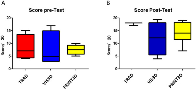
Results: Students of the whole cohort had significant better scores at the post test (mean score = 14.2) compared to the pretest (mean score = 7.9) (p = 0.0011). In the traditional and 3D printing groups, students had significant better scores in the post-test (mean scores = 17.7 and 14.2 respectively) than in the pre-test (mean scores = 8.2 and 7.5; p = 0.0247 and p = 0.0003 respectively). There was no significant difference between the pre and post-test scores for the 3D visualization group (mean score = 8.2 and 11.7 respectively) (p = 0.4347).
Conclusions: The knowledge of lung segment anatomy is poor among our medical students. Both traditional and 3D-printed teaching was shown effective. The contribution of 3D printed models would probably improve anatomy teaching among medical students. The introduction of this technology is instinctive and easy to use for both students and teachers. Furthermore, this technique was not particularly expensive to set up.

 求助内容:
求助内容: 应助结果提醒方式:


