Merridy J Lithgow, Jayishni N Maharaj, Andrew K Buldt, Shannon E Munteanu, Benjamin F Mentiplay, Hylton B Menz
{"title":"Lower Limb Kinematics of People With Midfoot Osteoarthritis During Level Walking and Stair Climbing.","authors":"Merridy J Lithgow, Jayishni N Maharaj, Andrew K Buldt, Shannon E Munteanu, Benjamin F Mentiplay, Hylton B Menz","doi":"10.1002/jfa2.70054","DOIUrl":null,"url":null,"abstract":"<p><strong>Background: </strong>Midfoot osteoarthritis (OA) affects one in eight people over 50, yet its impact on foot and lower limb kinematics remains poorly understood. This study compared foot and lower limb kinematics during level walking and stair climbing between people with and without symptomatic radiographic midfoot OA.</p><p><strong>Methods: </strong>Symptomatic radiographic midfoot OA was defined as midfoot pain in the last 4 weeks and radiographic OA in one or more midfoot joints. Cases aged ≥ 45 years were matched 1:1 for sex and age (± 5 years) to controls. A 10-camera motion analysis system was used to capture foot and lower limb kinematics during level walking and stair climbing, which were analysed with a validated multi-segmental lower limb model. Group differences were analysed using independent samples t-tests and effect sizes for discrete angles, whereas statistical parametric mapping compared kinematic patterns between groups.</p><p><strong>Results: </strong>We included 24 midfoot OA cases (mean age 64.4, SD 9.5) matched to 24 controls (mean age 65.2, SD 10.1). During level walking, people with midfoot OA walked slower and displayed absolute joint angles that showed less hip extension throughout stance, less knee flexion in early and late stance, less ankle dorsiflexion throughout stance (medium to large effects), greater subtalar pronation in late stance, and greater tarsometatarsal supination during early stance (medium effects). There were few differences during stair ascent and descent.</p><p><strong>Conclusion: </strong>People with midfoot OA walk slower and demonstrate medium to large differences in sagittal plane hip, knee, and ankle kinematics, and medium differences in subtalar and tarsometatarsal kinematics. These findings offer insights into the walking patterns of people with midfoot OA and the mechanisms that may contribute to or result from the condition. Prospective studies are needed to clarify the temporal relationship between these factors and midfoot OA development.</p>","PeriodicalId":49164,"journal":{"name":"Journal of Foot and Ankle Research","volume":"18 2","pages":"e70054"},"PeriodicalIF":2.2000,"publicationDate":"2025-06-01","publicationTypes":"Journal Article","fieldsOfStudy":null,"isOpenAccess":false,"openAccessPdf":"https://www.ncbi.nlm.nih.gov/pmc/articles/PMC12146581/pdf/","citationCount":"0","resultStr":null,"platform":"Semanticscholar","paperid":null,"PeriodicalName":"Journal of Foot and Ankle Research","FirstCategoryId":"3","ListUrlMain":"https://doi.org/10.1002/jfa2.70054","RegionNum":3,"RegionCategory":"医学","ArticlePicture":[],"TitleCN":null,"AbstractTextCN":null,"PMCID":null,"EPubDate":"","PubModel":"","JCR":"Q1","JCRName":"ORTHOPEDICS","Score":null,"Total":0}
引用次数: 0
Abstract
Background: Midfoot osteoarthritis (OA) affects one in eight people over 50, yet its impact on foot and lower limb kinematics remains poorly understood. This study compared foot and lower limb kinematics during level walking and stair climbing between people with and without symptomatic radiographic midfoot OA.
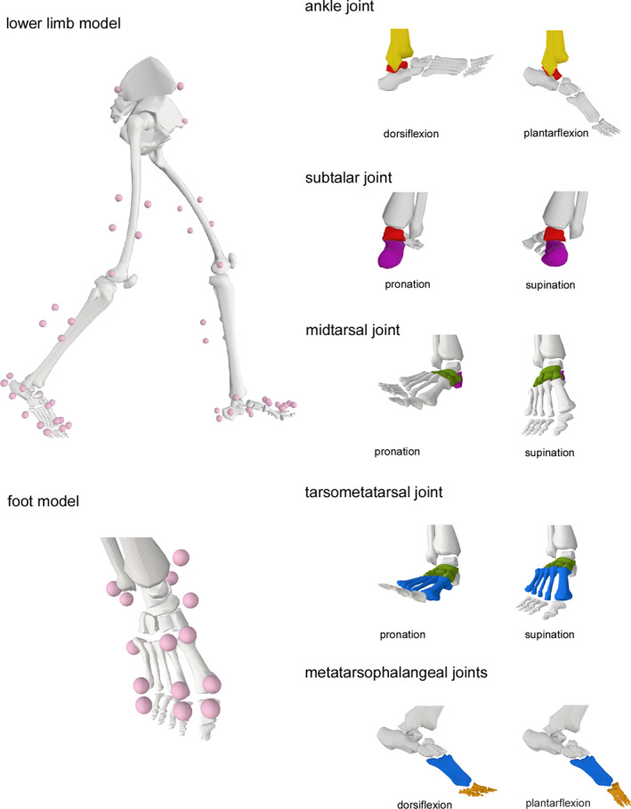
Methods: Symptomatic radiographic midfoot OA was defined as midfoot pain in the last 4 weeks and radiographic OA in one or more midfoot joints. Cases aged ≥ 45 years were matched 1:1 for sex and age (± 5 years) to controls. A 10-camera motion analysis system was used to capture foot and lower limb kinematics during level walking and stair climbing, which were analysed with a validated multi-segmental lower limb model. Group differences were analysed using independent samples t-tests and effect sizes for discrete angles, whereas statistical parametric mapping compared kinematic patterns between groups.
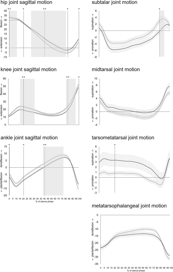
Results: We included 24 midfoot OA cases (mean age 64.4, SD 9.5) matched to 24 controls (mean age 65.2, SD 10.1). During level walking, people with midfoot OA walked slower and displayed absolute joint angles that showed less hip extension throughout stance, less knee flexion in early and late stance, less ankle dorsiflexion throughout stance (medium to large effects), greater subtalar pronation in late stance, and greater tarsometatarsal supination during early stance (medium effects). There were few differences during stair ascent and descent.
Conclusion: People with midfoot OA walk slower and demonstrate medium to large differences in sagittal plane hip, knee, and ankle kinematics, and medium differences in subtalar and tarsometatarsal kinematics. These findings offer insights into the walking patterns of people with midfoot OA and the mechanisms that may contribute to or result from the condition. Prospective studies are needed to clarify the temporal relationship between these factors and midfoot OA development.
期刊介绍:
Journal of Foot and Ankle Research, the official journal of the Australian Podiatry Association and The College of Podiatry (UK), is an open access journal that encompasses all aspects of policy, organisation, delivery and clinical practice related to the assessment, diagnosis, prevention and management of foot and ankle disorders.
Journal of Foot and Ankle Research covers a wide range of clinical subject areas, including diabetology, paediatrics, sports medicine, gerontology and geriatrics, foot surgery, physical therapy, dermatology, wound management, radiology, biomechanics and bioengineering, orthotics and prosthetics, as well the broad areas of epidemiology, policy, organisation and delivery of services related to foot and ankle care.
The journal encourages submissions from all health professionals who manage lower limb conditions, including podiatrists, nurses, physical therapists and physiotherapists, orthopaedists, manual therapists, medical specialists and general medical practitioners, as well as health service researchers concerned with foot and ankle care.
The Australian Podiatry Association and the College of Podiatry (UK) have reserve funds to cover the article-processing charge for manuscripts submitted by its members. Society members can email the appropriate contact at Australian Podiatry Association or The College of Podiatry to obtain the corresponding code to enter on submission.
 求助内容:
求助内容: 应助结果提醒方式:


