Farzaneh Ramezani, Sare Kamali, Ramin Mashoufi, Seyed Ali Ebrahimi, Maryam Soltan
{"title":"Sclerosing Angiomatoid Nodular Transformation of Spleen (SANTS); Case Report of a 12-Year-Old Patient.","authors":"Farzaneh Ramezani, Sare Kamali, Ramin Mashoufi, Seyed Ali Ebrahimi, Maryam Soltan","doi":"10.30699/ijp.2025.2034980.3332","DOIUrl":null,"url":null,"abstract":"<p><strong>Background: </strong>Sclerosing angiomatoid nodular transformation of the spleen (SANT) is a rare, benign vascular lesion predominantly described in adults. Pediatric cases are exceptionally uncommon and present a diagnostic challenge due to nonspecific clinical presentations and imaging findings.</p><p><strong>Case presentation: </strong>We report the case of a 12-year-old boy presenting with recurrent abdominal pain localized around the umbilicus, accompanied by intermittent nausea over a three-month period. Physical examination revealed mild tenderness without guarding. Laboratory findings were unremarkable. Abdominal ultrasound demonstrated a hypoechoic splenic lesion, further evaluated by multidetector computed tomography (MDCT), which revealed a heterogeneous hypodense mass in the spleen. The patient underwent partial laparoscopic splenectomy. Histopathological examination showed a nodular architecture with fibrous bands, capillary-like vascular channels lined by endothelial cells, and a lymphoplasmacytic infiltrate. Immunohistochemical staining was positive for CD31, CD34, and CD8, supporting the diagnosis of SANT.</p><p><strong>Conclusion: </strong>Although benign, SANT can mimic more aggressive splenic pathologies. This case underscores the importance of considering SANT in the differential diagnosis of splenic masses in pediatric patients and highlights the role of histopathology and immunohistochemistry in achieving a definitive diagnosis.</p>","PeriodicalId":38900,"journal":{"name":"Iranian Journal of Pathology","volume":"20 2","pages":"239-243"},"PeriodicalIF":0.0000,"publicationDate":"2025-01-01","publicationTypes":"Journal Article","fieldsOfStudy":null,"isOpenAccess":false,"openAccessPdf":"https://www.ncbi.nlm.nih.gov/pmc/articles/PMC12142018/pdf/","citationCount":"0","resultStr":null,"platform":"Semanticscholar","paperid":null,"PeriodicalName":"Iranian Journal of Pathology","FirstCategoryId":"1085","ListUrlMain":"https://doi.org/10.30699/ijp.2025.2034980.3332","RegionNum":0,"RegionCategory":null,"ArticlePicture":[],"TitleCN":null,"AbstractTextCN":null,"PMCID":null,"EPubDate":"2025/3/10 0:00:00","PubModel":"Epub","JCR":"Q3","JCRName":"Medicine","Score":null,"Total":0}
引用次数: 0
Abstract
Background: Sclerosing angiomatoid nodular transformation of the spleen (SANT) is a rare, benign vascular lesion predominantly described in adults. Pediatric cases are exceptionally uncommon and present a diagnostic challenge due to nonspecific clinical presentations and imaging findings.
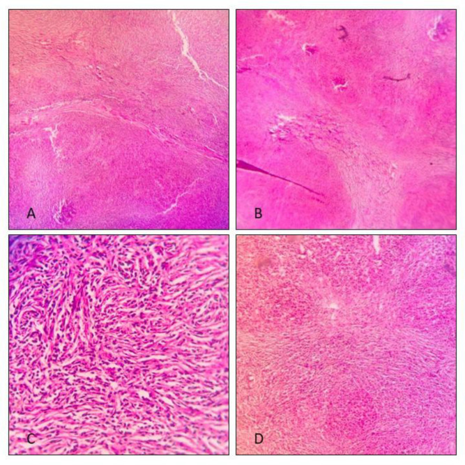
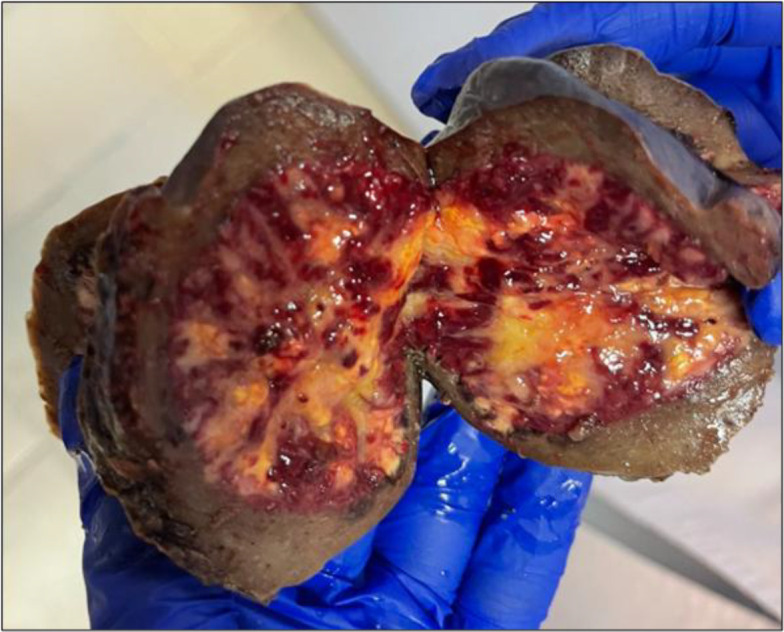
Case presentation: We report the case of a 12-year-old boy presenting with recurrent abdominal pain localized around the umbilicus, accompanied by intermittent nausea over a three-month period. Physical examination revealed mild tenderness without guarding. Laboratory findings were unremarkable. Abdominal ultrasound demonstrated a hypoechoic splenic lesion, further evaluated by multidetector computed tomography (MDCT), which revealed a heterogeneous hypodense mass in the spleen. The patient underwent partial laparoscopic splenectomy. Histopathological examination showed a nodular architecture with fibrous bands, capillary-like vascular channels lined by endothelial cells, and a lymphoplasmacytic infiltrate. Immunohistochemical staining was positive for CD31, CD34, and CD8, supporting the diagnosis of SANT.
Conclusion: Although benign, SANT can mimic more aggressive splenic pathologies. This case underscores the importance of considering SANT in the differential diagnosis of splenic masses in pediatric patients and highlights the role of histopathology and immunohistochemistry in achieving a definitive diagnosis.

 求助内容:
求助内容: 应助结果提醒方式:


