Corrado Campisi, Giovanni Giulietti, Carlo Alberto Artusi, Federico D'Agata, Giovanni Morana, Claudia Ledda, Elisa Montanaro, Mario Coriasco, Leonardo Lopiano, Marco Bozzali
{"title":"Clinical MRI to predict motor and non-motor effects of deep brain stimulation in Parkinson disease.","authors":"Corrado Campisi, Giovanni Giulietti, Carlo Alberto Artusi, Federico D'Agata, Giovanni Morana, Claudia Ledda, Elisa Montanaro, Mario Coriasco, Leonardo Lopiano, Marco Bozzali","doi":"10.1007/s11547-025-02025-8","DOIUrl":null,"url":null,"abstract":"<p><strong>Purpose: </strong>Subthalamic deep brain stimulation (STN-DBS) is a well-established intervention for advanced Parkinson's disease (PD). Routine neuroimaging can be used to estimate location and volume of activated tissue (VTA), by modeling the type of stimulator and stimulation parameters. We aimed here at developing a strategy based on clinical brain MRI scans to predict motor and non-motor outcomes of STN-DBS.</p><p><strong>Materials and methods: </strong>We included 25 consecutive patients with advanced PD eligible for STN-DBS. At baseline, patients underwent a comprehensive motor and cognitive/behavioral assessment, and conventional MRI. They underwent STN-DBS surgery, followed by a CT scan. Patients were reassessed 1 year later, while STN-DBS was active. Their neuroimaging data were used to calculate individual VTAs. The voxel-lesion-symptom-mapping (VLSM) toolbox, which allows to associate clinical variables with brain features of interest, was used to investigate associations between changes (in either direction) of motor, cognitive/behavioral scores between baseline and follow-up, and VTA subregions. Six newly enrolled patients were used to test the predictive value of this approach at a single subject level.</p><p><strong>Results: </strong>VLSM analysis (p values corrected for multiple comparisons < 0.05) identified specific VTA subclusters associated with improved bradykinesia, verbal fluency, and mood state, and some others associated with worsening of tremor, long-term memory, and apathy. When considering cognitive/behavioral changes, an effect of hemisphere lateralization was observed, with modulation of the right basal ganglia being associated with symptoms' worsening, and left-side modulation associated with improvements. VTA subclusters predictive for clinical changes were mostly located outside the STN, indicating the importance of networks over single nuclei simulation.</p><p><strong>Conclusion: </strong>This approach suggests a possible way to personalize surgical planning, DBS-implant choice, and stimulation programing in the framework of precision medicine.</p>","PeriodicalId":20817,"journal":{"name":"Radiologia Medica","volume":" ","pages":"1263-1274"},"PeriodicalIF":4.8000,"publicationDate":"2025-08-01","publicationTypes":"Journal Article","fieldsOfStudy":null,"isOpenAccess":false,"openAccessPdf":"https://www.ncbi.nlm.nih.gov/pmc/articles/PMC12367973/pdf/","citationCount":"0","resultStr":null,"platform":"Semanticscholar","paperid":null,"PeriodicalName":"Radiologia Medica","FirstCategoryId":"3","ListUrlMain":"https://doi.org/10.1007/s11547-025-02025-8","RegionNum":1,"RegionCategory":"医学","ArticlePicture":[],"TitleCN":null,"AbstractTextCN":null,"PMCID":null,"EPubDate":"2025/6/9 0:00:00","PubModel":"Epub","JCR":"Q1","JCRName":"RADIOLOGY, NUCLEAR MEDICINE & MEDICAL IMAGING","Score":null,"Total":0}
引用次数: 0
Abstract
Purpose: Subthalamic deep brain stimulation (STN-DBS) is a well-established intervention for advanced Parkinson's disease (PD). Routine neuroimaging can be used to estimate location and volume of activated tissue (VTA), by modeling the type of stimulator and stimulation parameters. We aimed here at developing a strategy based on clinical brain MRI scans to predict motor and non-motor outcomes of STN-DBS.
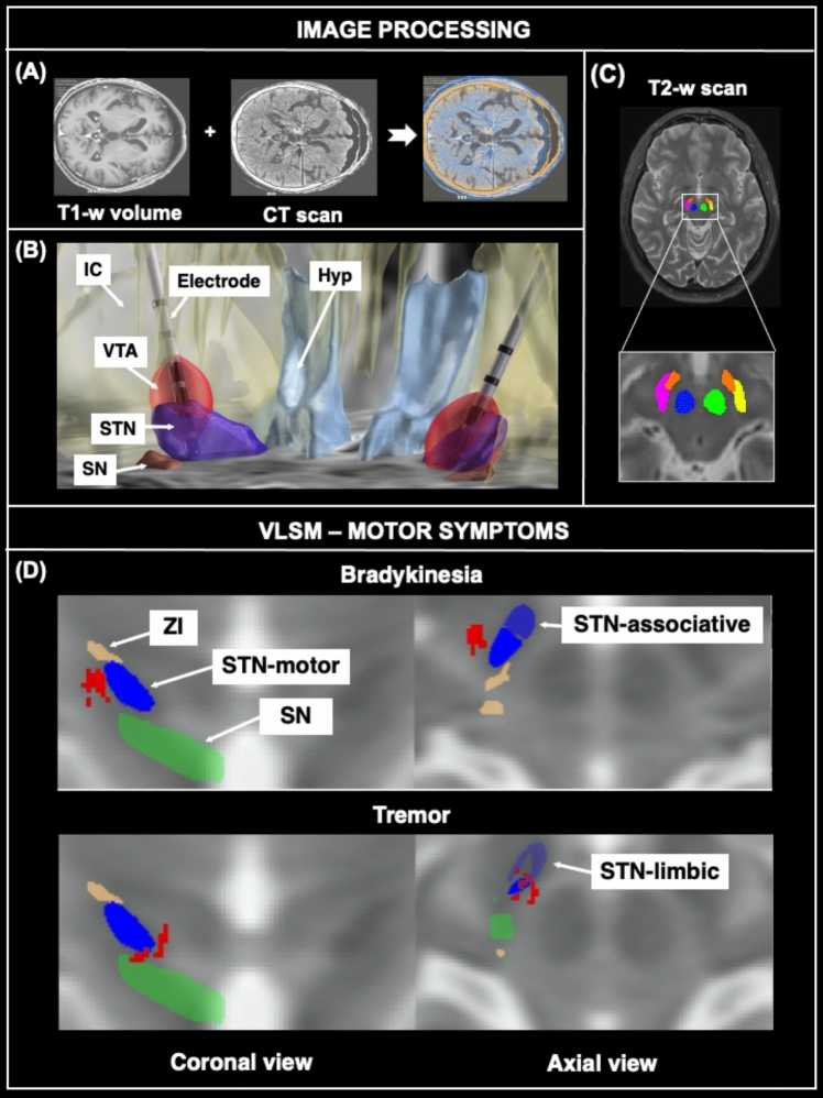
Materials and methods: We included 25 consecutive patients with advanced PD eligible for STN-DBS. At baseline, patients underwent a comprehensive motor and cognitive/behavioral assessment, and conventional MRI. They underwent STN-DBS surgery, followed by a CT scan. Patients were reassessed 1 year later, while STN-DBS was active. Their neuroimaging data were used to calculate individual VTAs. The voxel-lesion-symptom-mapping (VLSM) toolbox, which allows to associate clinical variables with brain features of interest, was used to investigate associations between changes (in either direction) of motor, cognitive/behavioral scores between baseline and follow-up, and VTA subregions. Six newly enrolled patients were used to test the predictive value of this approach at a single subject level.
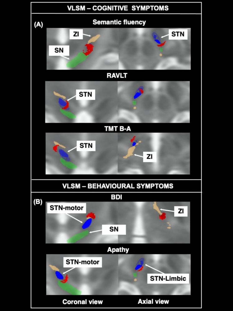
Results: VLSM analysis (p values corrected for multiple comparisons < 0.05) identified specific VTA subclusters associated with improved bradykinesia, verbal fluency, and mood state, and some others associated with worsening of tremor, long-term memory, and apathy. When considering cognitive/behavioral changes, an effect of hemisphere lateralization was observed, with modulation of the right basal ganglia being associated with symptoms' worsening, and left-side modulation associated with improvements. VTA subclusters predictive for clinical changes were mostly located outside the STN, indicating the importance of networks over single nuclei simulation.
Conclusion: This approach suggests a possible way to personalize surgical planning, DBS-implant choice, and stimulation programing in the framework of precision medicine.
期刊介绍:
Felice Perussia founded La radiologia medica in 1914. It is a peer-reviewed journal and serves as the official journal of the Italian Society of Medical and Interventional Radiology (SIRM). The primary purpose of the journal is to disseminate information related to Radiology, especially advancements in diagnostic imaging and related disciplines. La radiologia medica welcomes original research on both fundamental and clinical aspects of modern radiology, with a particular focus on diagnostic and interventional imaging techniques. It also covers topics such as radiotherapy, nuclear medicine, radiobiology, health physics, and artificial intelligence in the context of clinical implications. The journal includes various types of contributions such as original articles, review articles, editorials, short reports, and letters to the editor. With an esteemed Editorial Board and a selection of insightful reports, the journal is an indispensable resource for radiologists and professionals in related fields. Ultimately, La radiologia medica aims to serve as a platform for international collaboration and knowledge sharing within the radiological community.

 求助内容:
求助内容: 应助结果提醒方式:


