Artemisa Fernanda Moura Ferreira, Francisco de Assis Limeira-Júnior, José Jhenikártery Maia de Oliveira, Paulo Rogerio Ferreti Bonan, Marcelo Augusto Oliveira de Sales
{"title":"Imaging features of a brown tumor with extensive skull involvement: Relevance for dental radiology.","authors":"Artemisa Fernanda Moura Ferreira, Francisco de Assis Limeira-Júnior, José Jhenikártery Maia de Oliveira, Paulo Rogerio Ferreti Bonan, Marcelo Augusto Oliveira de Sales","doi":"10.4317/jced.62709","DOIUrl":null,"url":null,"abstract":"<p><p>This case report describes a rare, asymptomatic brown tumor in a patient with end-stage renal disease. The lesion was incidentally detected during a computed tomography (CT) scan of the paranasal sinuses, performed upon a dentist's recommendation to investigate maxillary sinusitis. CT imaging revealed an expansive osteolytic lesion with irregular margins and a ground-glass appearance involving the left side of the sphenoid and frontal sinuses. Subsequent magnetic resonance imaging (MRI) with multiplanar T1 and T2-weighted sequences without contrast demonstrated a solid tissue-like expansive lesion affecting the left frontal and sphenoid bones, mildly compressing adjacent cerebral parenchyma. Despite these findings, the patient remained asymptomatic. Conservative management, including pharmacological therapy with calcimimetics to control parathyroid hormone levels, was initiated. A follow-up MRI after five years showed lesion stability without significant changes. The patient later underwent a renal transplant, which effectively stabilized the bone disease and improved his quality of life. This case underscores the pivotal role of computed tomography (CT) in detecting incidental systemic skeletal changes and the indispensable importance of interdisciplinary collaboration in managing complex conditions in systemically compromised patients, where each professional's expertise is crucial for the patient's well-being. <b>Key words:</b>Sinusitis, Brown Tumor, Hyperparathyroidism, Multidetector Computed Tomography, Magnetic Resonance Imaging, Multidisciplinary Care Teams.</p>","PeriodicalId":15376,"journal":{"name":"Journal of Clinical and Experimental Dentistry","volume":"17 5","pages":"e604-e607"},"PeriodicalIF":0.0000,"publicationDate":"2025-05-01","publicationTypes":"Journal Article","fieldsOfStudy":null,"isOpenAccess":false,"openAccessPdf":"https://www.ncbi.nlm.nih.gov/pmc/articles/PMC12142367/pdf/","citationCount":"0","resultStr":null,"platform":"Semanticscholar","paperid":null,"PeriodicalName":"Journal of Clinical and Experimental Dentistry","FirstCategoryId":"1085","ListUrlMain":"https://doi.org/10.4317/jced.62709","RegionNum":0,"RegionCategory":null,"ArticlePicture":[],"TitleCN":null,"AbstractTextCN":null,"PMCID":null,"EPubDate":"","PubModel":"","JCR":"Q2","JCRName":"Dentistry","Score":null,"Total":0}
引用次数: 0
Abstract
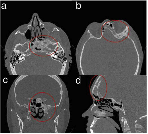
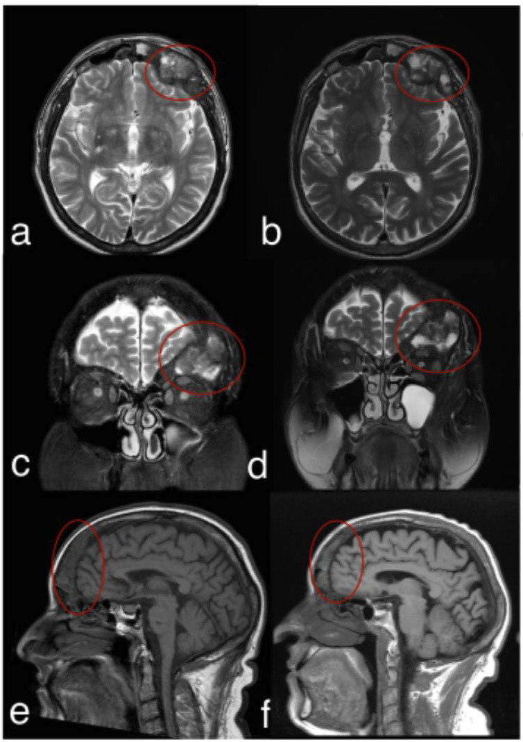
This case report describes a rare, asymptomatic brown tumor in a patient with end-stage renal disease. The lesion was incidentally detected during a computed tomography (CT) scan of the paranasal sinuses, performed upon a dentist's recommendation to investigate maxillary sinusitis. CT imaging revealed an expansive osteolytic lesion with irregular margins and a ground-glass appearance involving the left side of the sphenoid and frontal sinuses. Subsequent magnetic resonance imaging (MRI) with multiplanar T1 and T2-weighted sequences without contrast demonstrated a solid tissue-like expansive lesion affecting the left frontal and sphenoid bones, mildly compressing adjacent cerebral parenchyma. Despite these findings, the patient remained asymptomatic. Conservative management, including pharmacological therapy with calcimimetics to control parathyroid hormone levels, was initiated. A follow-up MRI after five years showed lesion stability without significant changes. The patient later underwent a renal transplant, which effectively stabilized the bone disease and improved his quality of life. This case underscores the pivotal role of computed tomography (CT) in detecting incidental systemic skeletal changes and the indispensable importance of interdisciplinary collaboration in managing complex conditions in systemically compromised patients, where each professional's expertise is crucial for the patient's well-being. Key words:Sinusitis, Brown Tumor, Hyperparathyroidism, Multidetector Computed Tomography, Magnetic Resonance Imaging, Multidisciplinary Care Teams.
期刊介绍:
Indexed in PUBMED, PubMed Central® (PMC) since 2012 and SCOPUSJournal of Clinical and Experimental Dentistry is an Open Access (free access on-line) - http://www.medicinaoral.com/odo/indice.htm. The aim of the Journal of Clinical and Experimental Dentistry is: - Periodontology - Community and Preventive Dentistry - Esthetic Dentistry - Biomaterials and Bioengineering in Dentistry - Operative Dentistry and Endodontics - Prosthetic Dentistry - Orthodontics - Oral Medicine and Pathology - Odontostomatology for the disabled or special patients - Oral Surgery

 求助内容:
求助内容: 应助结果提醒方式:


