Maria Eduarda Zen Biz, Jéssica Paola Salame, Gustavo Gumz Correia, Rafael Saviolo Moreira
{"title":"Variations in hepatic circulation: a study of 500 abdominal computed tomography scans.","authors":"Maria Eduarda Zen Biz, Jéssica Paola Salame, Gustavo Gumz Correia, Rafael Saviolo Moreira","doi":"10.1590/1677-5449.202401102","DOIUrl":null,"url":null,"abstract":"<p><strong>Background: </strong>Knowledge of the vascular anatomy of the liver and other abdominal organs helps surgeons improve preoperative planning, achieve greater surgical success, prevent complications, and reduce morbidity and mortality.</p><p><strong>Objectives: </strong>To report the prevalence of anatomical variation in the proper hepatic artery and portal vein observed through computed tomography.</p><p><strong>Methods: </strong>This retrospective study was based on 500 3-phase abdominal computed tomography scans. Variations in arterial anatomy were classified according to the Michels system (1966), while those in regarding portal vein anatomy were classified according to the Cheng system (1996).</p><p><strong>Results: </strong>A total of 31.2% of the cases showed variations in arterial vascularization, the most prevalent being type V (8.2%). No participants were identified with type X, and 0.4% could not be classified. A total of 21.8% showed variation in venous vascularization, with type IV being the most prevalent (8%).</p><p><strong>Conclusions: </strong>Medical knowledge of these variations and their prevalence is fundamental for the correct surgical management of upper abdomen pathologies and lower rates of postoperative complications. Variations not classified by previous trials should be categorized according to their clinical importance, and new studies should clarify national population patterns to reduce mortality rates from surgical procedures that involve these vessels.</p>","PeriodicalId":14814,"journal":{"name":"Jornal Vascular Brasileiro","volume":"24 ","pages":"e20240110"},"PeriodicalIF":1.0000,"publicationDate":"2025-05-30","publicationTypes":"Journal Article","fieldsOfStudy":null,"isOpenAccess":false,"openAccessPdf":"https://www.ncbi.nlm.nih.gov/pmc/articles/PMC12143226/pdf/","citationCount":"0","resultStr":null,"platform":"Semanticscholar","paperid":null,"PeriodicalName":"Jornal Vascular Brasileiro","FirstCategoryId":"1085","ListUrlMain":"https://doi.org/10.1590/1677-5449.202401102","RegionNum":0,"RegionCategory":null,"ArticlePicture":[],"TitleCN":null,"AbstractTextCN":null,"PMCID":null,"EPubDate":"2025/1/1 0:00:00","PubModel":"eCollection","JCR":"Q4","JCRName":"PERIPHERAL VASCULAR DISEASE","Score":null,"Total":0}
引用次数: 0
Abstract
Background: Knowledge of the vascular anatomy of the liver and other abdominal organs helps surgeons improve preoperative planning, achieve greater surgical success, prevent complications, and reduce morbidity and mortality.
Objectives: To report the prevalence of anatomical variation in the proper hepatic artery and portal vein observed through computed tomography.
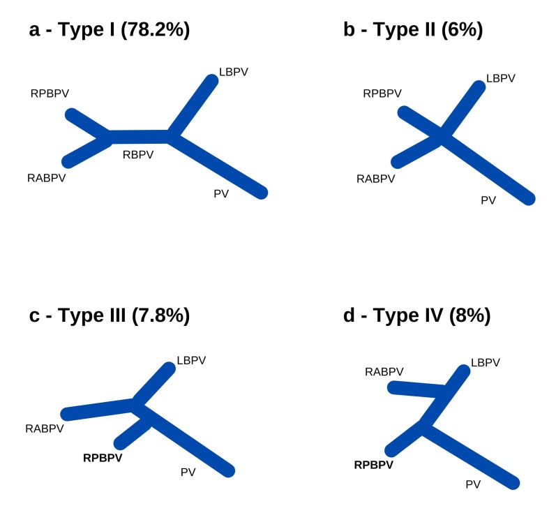
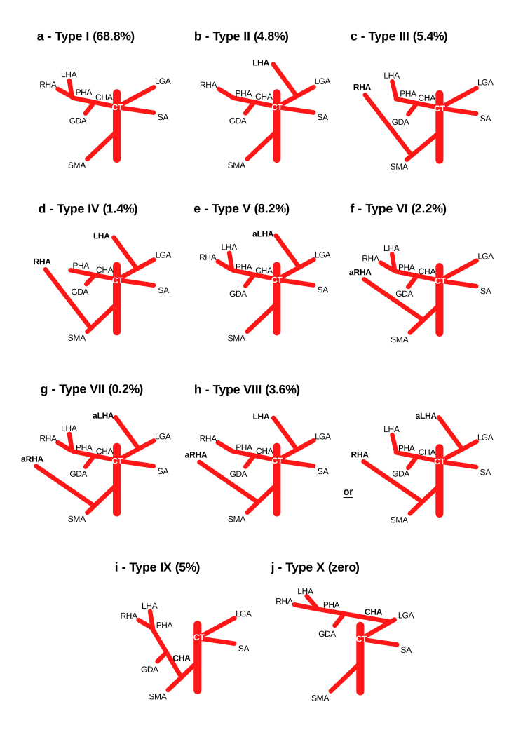
Methods: This retrospective study was based on 500 3-phase abdominal computed tomography scans. Variations in arterial anatomy were classified according to the Michels system (1966), while those in regarding portal vein anatomy were classified according to the Cheng system (1996).
Results: A total of 31.2% of the cases showed variations in arterial vascularization, the most prevalent being type V (8.2%). No participants were identified with type X, and 0.4% could not be classified. A total of 21.8% showed variation in venous vascularization, with type IV being the most prevalent (8%).
Conclusions: Medical knowledge of these variations and their prevalence is fundamental for the correct surgical management of upper abdomen pathologies and lower rates of postoperative complications. Variations not classified by previous trials should be categorized according to their clinical importance, and new studies should clarify national population patterns to reduce mortality rates from surgical procedures that involve these vessels.
期刊介绍:
The Jornal Vascular Brasileiro is editated and published quaterly to select and disseminate high-quality scientific contents concerning original research, novel surgical and diagnostic techniques, and clinical observations in the field of vascular surgery, angiology, and endovascular surgery. Its abbreviated title is J. Vasc. Bras., which should be used in bibliographies, footnotes and bibliographical references and strips.


 求助内容:
求助内容: 应助结果提醒方式:


