Marc Anton Fuessinger, Maximilian Frederik Russe, Leonard Simon Brandenburg, Marc Christian Metzger, Johannes Schulze, Stefan Schlager, Jonas Wuester, Wiebke Semper-Hogg
{"title":"Optimization of the size and location of the FOVs for CBCT capture of impacted lower third molars.","authors":"Marc Anton Fuessinger, Maximilian Frederik Russe, Leonard Simon Brandenburg, Marc Christian Metzger, Johannes Schulze, Stefan Schlager, Jonas Wuester, Wiebke Semper-Hogg","doi":"10.1186/s13005-025-00518-5","DOIUrl":null,"url":null,"abstract":"<p><strong>Background: </strong>Cone beam computed tomography (CBCT) is an established diagnostic tool for impacted wisdom teeth (third molars (3 M)) in proximity of the mandibular nerve canal. This study aims to define the minimum field-of-view (FOV) size and its localization to reduce radiation exposure. As reference, the chin rest of the CBCT device was used.</p><p><strong>Methods: </strong>Three-dimensional CBCT data sets were used to analyze the bilateral positions and dimensions of the wisdom teeth. A total of 215 wisdom teeth from a study population with a mean age of 21 years, including data from 82 male and 58 female patients, were mapped. By transformation into a common coordinate space using the device's chin rest as a joint denominator, the optimal size and location for uni- and bilateral capture of the wisdom teeth were determined, for both best-case and worst-case scenarios with regard to patient positioning.</p><p><strong>Results: </strong>The minimal FOVs for the lower 3 M capture were H 23.5 mm × R 35.4 mm in the best-case scenario assuming optimal patient positioning and H 35.4 mm × R 36.6 mm in the worst-case scenario with rotational deviation along the transversal axis. For the upper 3 M, the minimal FOVs were H 29.9 mm × R 29.2 mm in the best-case scenario and H 38.6 mm × R 35.6 mm in the worst-case scenario. Unilateral capture of both the upper and lower 3 M required FOV dimensions of H 51.7 mm × R 39.8 mm and H 44.8 mm × R 36.8 mm, respectively. For bilateral capture of all four 3 M, the best-case FOV was H 44.8 mm × R 84.8 mm and the worst-case FOV was H 51.7 mm × R 85.6 mm.</p><p><strong>Discussion: </strong>This research provides indication-specific FOVs for uni- and bilateral imaging of the upper and lower 3 M. Taking into account optimal clinical practices for CBCT imaging, this study aims to propose clinically feasible FOV dimensions while meeting the technical specifications of commonly used CBCT devices. Clinical application of the results may help reduce radiation exposure of patients receiving CBCT imaging of the wisdom teeth. Transfer of the present results to other CBCT devices requires further research.</p><p><strong>Trial registration: </strong>The study is registered in the German Trial Register with the number DRKS00026149, 2024/02/21.</p>","PeriodicalId":12994,"journal":{"name":"Head & Face Medicine","volume":"21 1","pages":"45"},"PeriodicalIF":2.4000,"publicationDate":"2025-06-07","publicationTypes":"Journal Article","fieldsOfStudy":null,"isOpenAccess":false,"openAccessPdf":"https://www.ncbi.nlm.nih.gov/pmc/articles/PMC12144699/pdf/","citationCount":"0","resultStr":null,"platform":"Semanticscholar","paperid":null,"PeriodicalName":"Head & Face Medicine","FirstCategoryId":"3","ListUrlMain":"https://doi.org/10.1186/s13005-025-00518-5","RegionNum":2,"RegionCategory":"医学","ArticlePicture":[],"TitleCN":null,"AbstractTextCN":null,"PMCID":null,"EPubDate":"","PubModel":"","JCR":"Q2","JCRName":"DENTISTRY, ORAL SURGERY & MEDICINE","Score":null,"Total":0}
引用次数: 0
Abstract
Background: Cone beam computed tomography (CBCT) is an established diagnostic tool for impacted wisdom teeth (third molars (3 M)) in proximity of the mandibular nerve canal. This study aims to define the minimum field-of-view (FOV) size and its localization to reduce radiation exposure. As reference, the chin rest of the CBCT device was used.
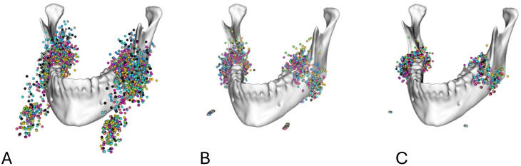
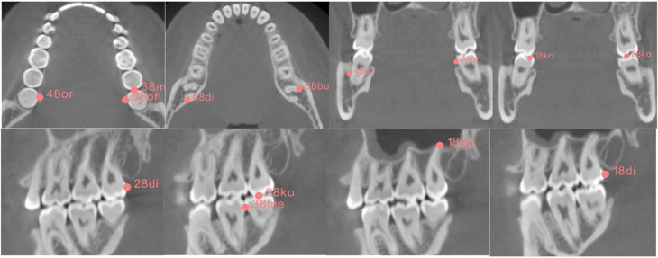
Methods: Three-dimensional CBCT data sets were used to analyze the bilateral positions and dimensions of the wisdom teeth. A total of 215 wisdom teeth from a study population with a mean age of 21 years, including data from 82 male and 58 female patients, were mapped. By transformation into a common coordinate space using the device's chin rest as a joint denominator, the optimal size and location for uni- and bilateral capture of the wisdom teeth were determined, for both best-case and worst-case scenarios with regard to patient positioning.
Results: The minimal FOVs for the lower 3 M capture were H 23.5 mm × R 35.4 mm in the best-case scenario assuming optimal patient positioning and H 35.4 mm × R 36.6 mm in the worst-case scenario with rotational deviation along the transversal axis. For the upper 3 M, the minimal FOVs were H 29.9 mm × R 29.2 mm in the best-case scenario and H 38.6 mm × R 35.6 mm in the worst-case scenario. Unilateral capture of both the upper and lower 3 M required FOV dimensions of H 51.7 mm × R 39.8 mm and H 44.8 mm × R 36.8 mm, respectively. For bilateral capture of all four 3 M, the best-case FOV was H 44.8 mm × R 84.8 mm and the worst-case FOV was H 51.7 mm × R 85.6 mm.
Discussion: This research provides indication-specific FOVs for uni- and bilateral imaging of the upper and lower 3 M. Taking into account optimal clinical practices for CBCT imaging, this study aims to propose clinically feasible FOV dimensions while meeting the technical specifications of commonly used CBCT devices. Clinical application of the results may help reduce radiation exposure of patients receiving CBCT imaging of the wisdom teeth. Transfer of the present results to other CBCT devices requires further research.
Trial registration: The study is registered in the German Trial Register with the number DRKS00026149, 2024/02/21.
背景:圆锥束计算机断层扫描(CBCT)是下颌神经管附近阻生智齿(第三磨牙(3 M))的诊断工具。本研究旨在确定最小视场(FOV)大小及其定位,以减少辐射暴露。作为参考,采用CBCT装置的下巴托。方法:采用三维CBCT数据集对双侧智齿位置和尺寸进行分析。研究人员对平均年龄为21岁的研究人群中的215颗智齿进行了绘制,其中包括82名男性和58名女性患者的数据。通过变换到一个共同的坐标空间,以设备的下巴托为联合分母,确定了单侧和双侧智齿捕获的最佳尺寸和位置,同时考虑了患者定位的最佳情况和最坏情况。结果:在最佳患者体位的最佳情况下,下3m捕获的最小fov为H 23.5 mm × R 35.4 mm;在沿横向轴旋转偏差的最坏情况下,h35.4 mm × R 36.6 mm。在高3米的情况下,最佳情况下的最小视场为H 29.9 mm × R 29.2 mm,最差情况下为H 38.6 mm × R 35.6 mm。单侧捕获上、下3 M所需的视场尺寸分别为高51.7 mm × R 39.8 mm和高44.8 mm × R 36.8 mm。对于所有4个3 M的双侧捕获,最佳情况FOV为H 44.8 mm × R 84.8 mm,最坏情况FOV为H 51.7 mm × R 85.6 mm。讨论:本研究为上下3 M的单侧和双侧成像提供了特定适应症的FOV。考虑到CBCT成像的最佳临床实践,本研究旨在提出临床可行的FOV尺寸,同时满足常用CBCT设备的技术规范。本研究结果的临床应用将有助于减少患者接受智慧牙CBCT成像时的辐射暴露。将目前的结果转移到其他CBCT设备需要进一步的研究。试验注册:该研究已在德国试验注册中注册,编号为DRKS00026149, 20124/02/21。
期刊介绍:
Head & Face Medicine is a multidisciplinary open access journal that publishes basic and clinical research concerning all aspects of cranial, facial and oral conditions.
The journal covers all aspects of cranial, facial and oral diseases and their management. It has been designed as a multidisciplinary journal for clinicians and researchers involved in the diagnostic and therapeutic aspects of diseases which affect the human head and face. The journal is wide-ranging, covering the development, aetiology, epidemiology and therapy of head and face diseases to the basic science that underlies these diseases. Management of head and face diseases includes all aspects of surgical and non-surgical treatments including psychopharmacological therapies.
 求助内容:
求助内容: 应助结果提醒方式:


