Enhancing biliary tract cancer diagnosis using AI-driven 3D optical diffraction tomography
IF 4.3
3区 生物学
Q1 BIOCHEMICAL RESEARCH METHODS
引用次数: 0
Abstract
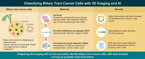
Biliary tract cancer is associated with distinct metabolic alterations, particularly in lipid metabolism. This study aimed to classify biliary tract cancer cells automatically based on lipid droplet (LD) characteristics using three-dimensional (3D) optical diffraction tomography (ODT) combined with convolutional neural networks (CNNs). Human biliary tract cancer cell lines (SNU1196, SNU308, and SNU478) and a normal cholangiocyte cell line (H69) were cultured to evaluate the LD volume, mass, and count. We generated 3D refractive index tomograms and developed a CNN-based diagnostic system for automated classification. The biliary tract cancer cells exhibited significantly increased LD volume, mass, and count compared with those of normal cholangiocytes, reflecting distinct metabolic profiles. The EfficientNet-b3 model achieved an area under the curve (AUC) of 0.982 and an accuracy of 93.79%. Incorporating LD metadata, such as volume and dry mass, improved performance, yielding an AUC of 0.997 and an accuracy of 97.94%. Combining LD metadata with multi-view score fusion enhanced diagnostic performance (AUC: 0.999, accuracy: 98.61%). Further, LayerCAM analysis revealed that the model focused on LD-rich cytoplasmic regions, thereby aligning with known metabolic phenotypes. Overall, our findings demonstrate the diagnostic potential of LD characteristics and support the clinical utility of 3D ODT combined with deep learning for early detection of biliary tract cancer and future multimodal applications.
人工智能驱动的三维光学衍射断层扫描增强胆道癌诊断。
胆道癌与明显的代谢改变有关,尤其是脂质代谢。本研究旨在利用三维光学衍射断层扫描(ODT)结合卷积神经网络(cnn),基于脂滴(LD)特征对胆道癌细胞进行自动分类。培养人胆道癌细胞系(SNU1196、SNU308和SNU478)和正常胆管细胞系(H69),评估LD的体积、质量和计数。我们生成了3D折射率断层图,并开发了一个基于cnn的自动分类诊断系统。与正常胆管细胞相比,胆道癌细胞的LD体积、质量和计数明显增加,反映了不同的代谢特征。effentnet -b3模型的曲线下面积(AUC)为0.982,准确度为93.79%。结合LD元数据,如体积和干质量,提高了性能,AUC为0.997,准确率为97.94%。结合LD元数据和多视图评分融合提高了诊断性能(AUC: 0.999,准确率:98.61%)。此外,LayerCAM分析显示,该模型专注于富含ld的细胞质区域,从而与已知的代谢表型一致。总的来说,我们的研究结果证明了LD特征的诊断潜力,并支持3D ODT结合深度学习在胆道癌早期检测中的临床应用,以及未来的多模式应用。
本文章由计算机程序翻译,如有差异,请以英文原文为准。
求助全文
约1分钟内获得全文
求助全文
来源期刊
Methods
生物-生化研究方法
CiteScore
9.80
自引率
2.10%
发文量
222
审稿时长
11.3 weeks
期刊介绍:
Methods focuses on rapidly developing techniques in the experimental biological and medical sciences.
Each topical issue, organized by a guest editor who is an expert in the area covered, consists solely of invited quality articles by specialist authors, many of them reviews. Issues are devoted to specific technical approaches with emphasis on clear detailed descriptions of protocols that allow them to be reproduced easily. The background information provided enables researchers to understand the principles underlying the methods; other helpful sections include comparisons of alternative methods giving the advantages and disadvantages of particular methods, guidance on avoiding potential pitfalls, and suggestions for troubleshooting.
 求助内容:
求助内容: 应助结果提醒方式:


