Amanda Rodriguez, Oliver Kripfgans, Fabiana Aellos, Diego Velasquez, Alejandra Baltazar, Hsun-Liang Chan
{"title":"Non-invasive and quantitative methods for assessment of blood flow in periodontal and oral soft tissues: a systematic review.","authors":"Amanda Rodriguez, Oliver Kripfgans, Fabiana Aellos, Diego Velasquez, Alejandra Baltazar, Hsun-Liang Chan","doi":"10.3389/fdmed.2025.1587821","DOIUrl":null,"url":null,"abstract":"<p><strong>Objectives: </strong>Understanding the available methods to study blood flow in the oral cavity can enhance knowledge of research methodology on periodontal circulation related to disease initiation and progression as well as wound healing. This study aims to systematically review non-invasive techniques that allow for the assessment of oral tissue perfusion in clinical and pre-clinical studies.</p><p><strong>Methods: </strong>A complete electronic literature search in 5 databases (NLM PubMed, Embase, EBSCOhost CINAHL, EBSCOhost Dentistry and Oral Sciences Source, and Wiley Cochrane Central Register of Controlled Trials) was conducted by two reviewers. The search terms included gingival blood flow, tissue perfusion, imaging perfusion, soft tissue perfusion, diagnostic, vascularization, soft tissue, and microvascularization. The focused question is: What are the available non-invasive and quantitative imaging techniques used to evaluate oral and periodontal tissue perfusion?</p><p><strong>Results: </strong>A total of 79 articles were included for qualitative analysis. Various methods were identified, including Laser Doppler Flowmetry (LDF), Laser Speckle Contrast Imaging (LSCI), Spectral Imaging Methods (such as Diffuse Reflectance Spectroscopy), Ultrasound (US), Intravital Video Microscopy, and Oral Videocapillaroscopy. LDF is the most applied to estimate blood flow in a small focal area for the study of periodontal diseases and oral wound healing, among other indications. LSCI, providing surrogate superficial blood flow values in a 2-dimensional, larger field-of-view, has been used for similar reasons. The use of cross-sectional ultrasound is on a rise to record blood velocity and blood volume using color flow and color power modes, respectively. Comparisons of the available technologies revealed their strengths and limitations related to their spatial resolution, sensitivity, reliability, accuracy, invasiveness, dependence of (image) data in the field of view relative to probe positioning and angulation, and safety. The ideal features of such a device pertinent to probe geometry, data acquisition, recording, and infection control needs were also discussed.</p><p><strong>Conclusions: </strong>A few imaging technologies have been identified in the literature to study blood flow in the oral cavity. These methods could potentially augment our ability to diagnose oral diseases and monitor wound healing objectively and timely. In combination, these could potentially enhance treatment outcomes significantly.</p>","PeriodicalId":73077,"journal":{"name":"Frontiers in dental medicine","volume":"6 ","pages":"1587821"},"PeriodicalIF":1.8000,"publicationDate":"2025-05-22","publicationTypes":"Journal Article","fieldsOfStudy":null,"isOpenAccess":false,"openAccessPdf":"https://www.ncbi.nlm.nih.gov/pmc/articles/PMC12137316/pdf/","citationCount":"0","resultStr":null,"platform":"Semanticscholar","paperid":null,"PeriodicalName":"Frontiers in dental medicine","FirstCategoryId":"1085","ListUrlMain":"https://doi.org/10.3389/fdmed.2025.1587821","RegionNum":0,"RegionCategory":null,"ArticlePicture":[],"TitleCN":null,"AbstractTextCN":null,"PMCID":null,"EPubDate":"2025/1/1 0:00:00","PubModel":"eCollection","JCR":"Q3","JCRName":"DENTISTRY, ORAL SURGERY & MEDICINE","Score":null,"Total":0}
引用次数: 0
Abstract
Objectives: Understanding the available methods to study blood flow in the oral cavity can enhance knowledge of research methodology on periodontal circulation related to disease initiation and progression as well as wound healing. This study aims to systematically review non-invasive techniques that allow for the assessment of oral tissue perfusion in clinical and pre-clinical studies.
Methods: A complete electronic literature search in 5 databases (NLM PubMed, Embase, EBSCOhost CINAHL, EBSCOhost Dentistry and Oral Sciences Source, and Wiley Cochrane Central Register of Controlled Trials) was conducted by two reviewers. The search terms included gingival blood flow, tissue perfusion, imaging perfusion, soft tissue perfusion, diagnostic, vascularization, soft tissue, and microvascularization. The focused question is: What are the available non-invasive and quantitative imaging techniques used to evaluate oral and periodontal tissue perfusion?
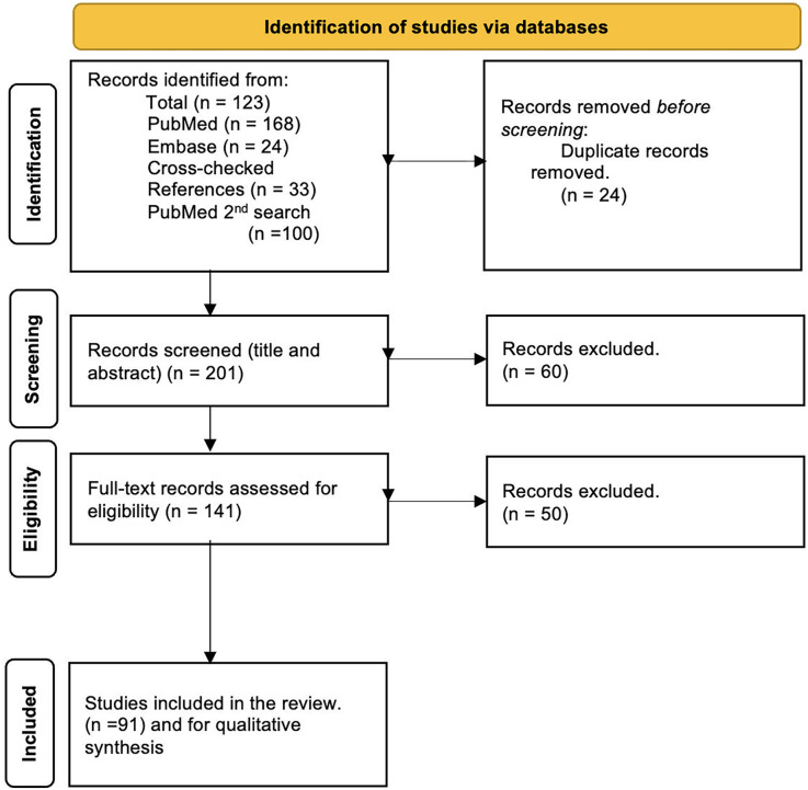
Results: A total of 79 articles were included for qualitative analysis. Various methods were identified, including Laser Doppler Flowmetry (LDF), Laser Speckle Contrast Imaging (LSCI), Spectral Imaging Methods (such as Diffuse Reflectance Spectroscopy), Ultrasound (US), Intravital Video Microscopy, and Oral Videocapillaroscopy. LDF is the most applied to estimate blood flow in a small focal area for the study of periodontal diseases and oral wound healing, among other indications. LSCI, providing surrogate superficial blood flow values in a 2-dimensional, larger field-of-view, has been used for similar reasons. The use of cross-sectional ultrasound is on a rise to record blood velocity and blood volume using color flow and color power modes, respectively. Comparisons of the available technologies revealed their strengths and limitations related to their spatial resolution, sensitivity, reliability, accuracy, invasiveness, dependence of (image) data in the field of view relative to probe positioning and angulation, and safety. The ideal features of such a device pertinent to probe geometry, data acquisition, recording, and infection control needs were also discussed.
Conclusions: A few imaging technologies have been identified in the literature to study blood flow in the oral cavity. These methods could potentially augment our ability to diagnose oral diseases and monitor wound healing objectively and timely. In combination, these could potentially enhance treatment outcomes significantly.
目的:了解现有的口腔血流研究方法,可以提高对牙周循环与疾病发生、发展和伤口愈合相关研究方法的认识。本研究旨在系统回顾临床和临床前研究中用于评估口腔组织灌注的非侵入性技术。方法:由2位审评员对5个数据库(NLM PubMed、Embase、EBSCOhost CINAHL、EBSCOhost Dentistry and Oral Sciences Source和Wiley Cochrane Central Register of Controlled Trials)进行完整的电子文献检索。搜索词包括牙龈血流、组织灌注、成像灌注、软组织灌注、诊断、血管化、软组织和微血管化。重点问题是:有哪些可用的非侵入性和定量成像技术用于评估口腔和牙周组织灌注?结果:共纳入79篇文献进行定性分析。确定了各种方法,包括激光多普勒血流法(LDF),激光散斑对比成像(LSCI),光谱成像方法(如漫反射光谱),超声(US),活体视频显微镜和口腔视频毛细血管镜检查。除其他适应症外,LDF最适用于估计牙周病和口腔伤口愈合研究的小焦点区域的血流量。LSCI在二维、更大的视野范围内提供替代的浅表血流值,也被用于类似的原因。使用横断面超声分别使用彩色流量和彩色功率模式记录血流速度和血容量的情况正在增加。通过对现有技术的比较,揭示了它们在空间分辨率、灵敏度、可靠性、准确性、侵入性、相对于探针定位和角度的视场(图像)数据依赖性以及安全性方面的优势和局限性。本文还讨论了该装置在探头几何形状、数据采集、记录和感染控制等方面的理想特性。结论:文献中已经确定了几种用于研究口腔血流的成像技术。这些方法有可能增强我们客观、及时地诊断口腔疾病和监测伤口愈合的能力。综合起来,这些可能会显著提高治疗效果。

 求助内容:
求助内容: 应助结果提醒方式:


