Pan Yu, Nan Hong, Genwang Wang, Shuqiang Chen, Zhipeng Zhao
{"title":"Hydrogen gas therapy: A promising approach for sepsis management post-burn injury by modulating inflammation, oxidative stress, and wound healing.","authors":"Pan Yu, Nan Hong, Genwang Wang, Shuqiang Chen, Zhipeng Zhao","doi":"10.25259/Cytojournal_253_2024","DOIUrl":null,"url":null,"abstract":"<p><strong>Objective: </strong>Burns refers to a severe form of trauma that often leads to localized and systemic inflammatory responses, oxidative stress, and immune dysfunction. Patients with severe burns are highly susceptible to the development of postburn sepsis, a condition influenced by multiple factors, such as bacterial infection of the burn wound, alterations in immune status, and excessive release of inflammatory mediators. This study aimed to investigate the mechanisms by which hydrogen gas treatment exerts its effects on postburn sepsis, with a focus on its influence on inflammatory responses, oxidative stress, and wound healing.</p><p><strong>Material and methods: </strong>This work employed <i>in vitro</i> assays with Sprague-Dawley (SD) rat skin fibroblasts (RSFs) to assess the effects of burn serum and hydrogen gas on cell proliferation through methylthiazolyldiphenyltetrazolium bromide assays and on apoptosis through flow cytometry with Annexin V-fluorescein isothiocyanate/propidium iodide staining. In addition, an enzyme-linked immunosorbent assay was performed to quantify inflammatory cytokines and oxidative stress markers in fibroblasts treated with burn serum. Western blotting (WB) analysis was conducted to investigate signaling pathway modulation. The severe burn sepsis models of SD rats were segregated into three experimental groups: a healthy normal control group, a burn sepsis control group, and a burn sepsis + hydrogen gas (2%) treatment group. Wound healing was monitored, with wound contraction rates recorded and histological assessments conducted using hematoxylin and eosin and Masson's trichrome staining to evaluate tissue repair and collagen deposition.</p><p><strong>Results: </strong><i>In vitro</i> assays showed that burn serum reduced fibroblast proliferation and increased apoptosis (<i>P</i> < 0.01), which hydrogen gas mitigated by rescuing cell viability and reducing apoptosis (<i>P</i> < 0.01). Enzyme-linked immunosorbent assay revealed burn serum-induced increases in the levels of inflammatory cytokines and oxidative stress markers, with decreases in antioxidant enzymes (<i>P</i> < 0.01), which hydrogen gas reversed (<i>P</i> < 0.05). WB analysis suggested hydrogen gas's anti-inflammatory and proliferative effects by modulating signaling pathways (<i>P</i> < 0.01). <i>In vivo</i>, hydrogen gas treatment considerably improved wound healing, with accelerated contraction and enhanced collagen deposition. Plasma and skin tissue analyses indicated systemic and local anti-inflammatory and antioxidant effects from hydrogen gas.</p><p><strong>Conclusion: </strong>Hydrogen gas treatment demonstrates potential therapeutic efficacy in the management of postburn sepsis by modulating inflammatory responses, reducing oxidative stress, and promoting wound healing. These findings provide scientific evidence supporting hydrogen gas as an adjunctive treatment strategy for postburn sepsis.</p>","PeriodicalId":49082,"journal":{"name":"Cytojournal","volume":"22 ","pages":"46"},"PeriodicalIF":3.1000,"publicationDate":"2025-04-25","publicationTypes":"Journal Article","fieldsOfStudy":null,"isOpenAccess":false,"openAccessPdf":"https://www.ncbi.nlm.nih.gov/pmc/articles/PMC12134856/pdf/","citationCount":"0","resultStr":null,"platform":"Semanticscholar","paperid":null,"PeriodicalName":"Cytojournal","FirstCategoryId":"3","ListUrlMain":"https://doi.org/10.25259/Cytojournal_253_2024","RegionNum":4,"RegionCategory":"医学","ArticlePicture":[],"TitleCN":null,"AbstractTextCN":null,"PMCID":null,"EPubDate":"2025/1/1 0:00:00","PubModel":"eCollection","JCR":"Q2","JCRName":"PATHOLOGY","Score":null,"Total":0}
引用次数: 0
Abstract
Objective: Burns refers to a severe form of trauma that often leads to localized and systemic inflammatory responses, oxidative stress, and immune dysfunction. Patients with severe burns are highly susceptible to the development of postburn sepsis, a condition influenced by multiple factors, such as bacterial infection of the burn wound, alterations in immune status, and excessive release of inflammatory mediators. This study aimed to investigate the mechanisms by which hydrogen gas treatment exerts its effects on postburn sepsis, with a focus on its influence on inflammatory responses, oxidative stress, and wound healing.
Material and methods: This work employed in vitro assays with Sprague-Dawley (SD) rat skin fibroblasts (RSFs) to assess the effects of burn serum and hydrogen gas on cell proliferation through methylthiazolyldiphenyltetrazolium bromide assays and on apoptosis through flow cytometry with Annexin V-fluorescein isothiocyanate/propidium iodide staining. In addition, an enzyme-linked immunosorbent assay was performed to quantify inflammatory cytokines and oxidative stress markers in fibroblasts treated with burn serum. Western blotting (WB) analysis was conducted to investigate signaling pathway modulation. The severe burn sepsis models of SD rats were segregated into three experimental groups: a healthy normal control group, a burn sepsis control group, and a burn sepsis + hydrogen gas (2%) treatment group. Wound healing was monitored, with wound contraction rates recorded and histological assessments conducted using hematoxylin and eosin and Masson's trichrome staining to evaluate tissue repair and collagen deposition.
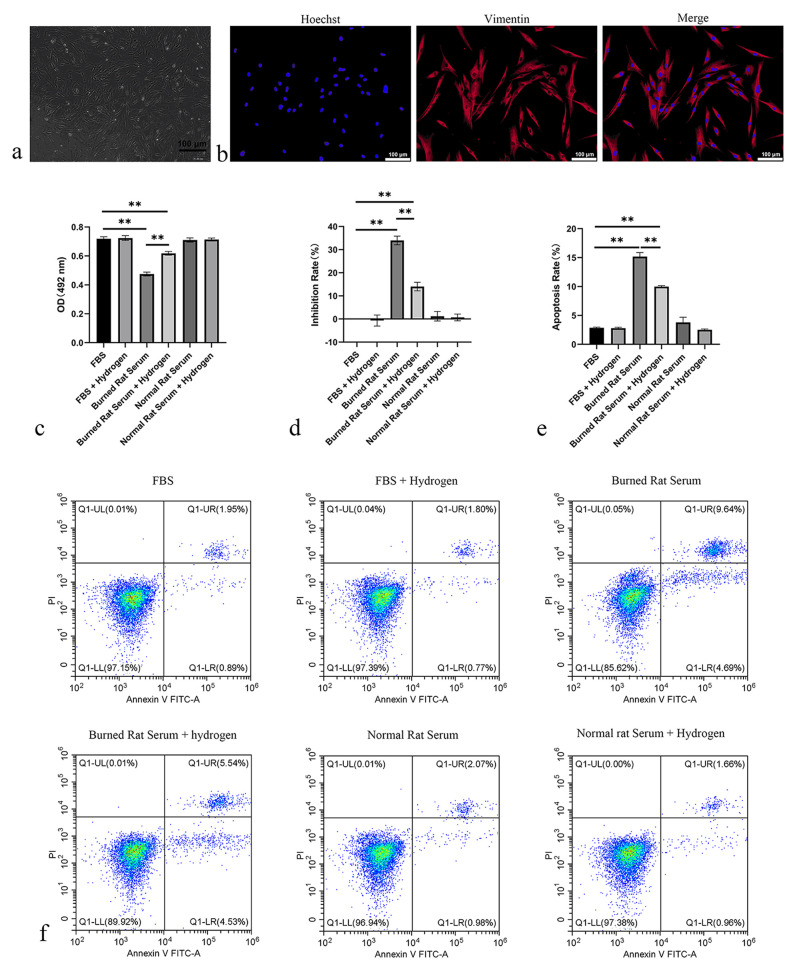
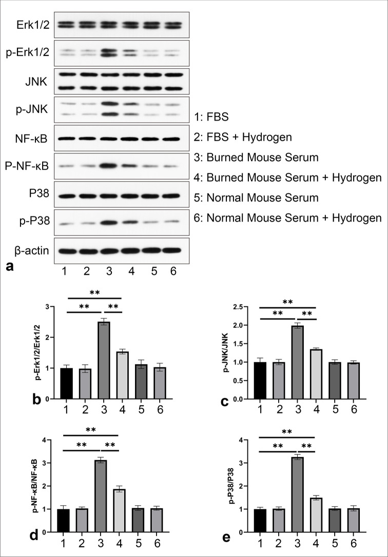
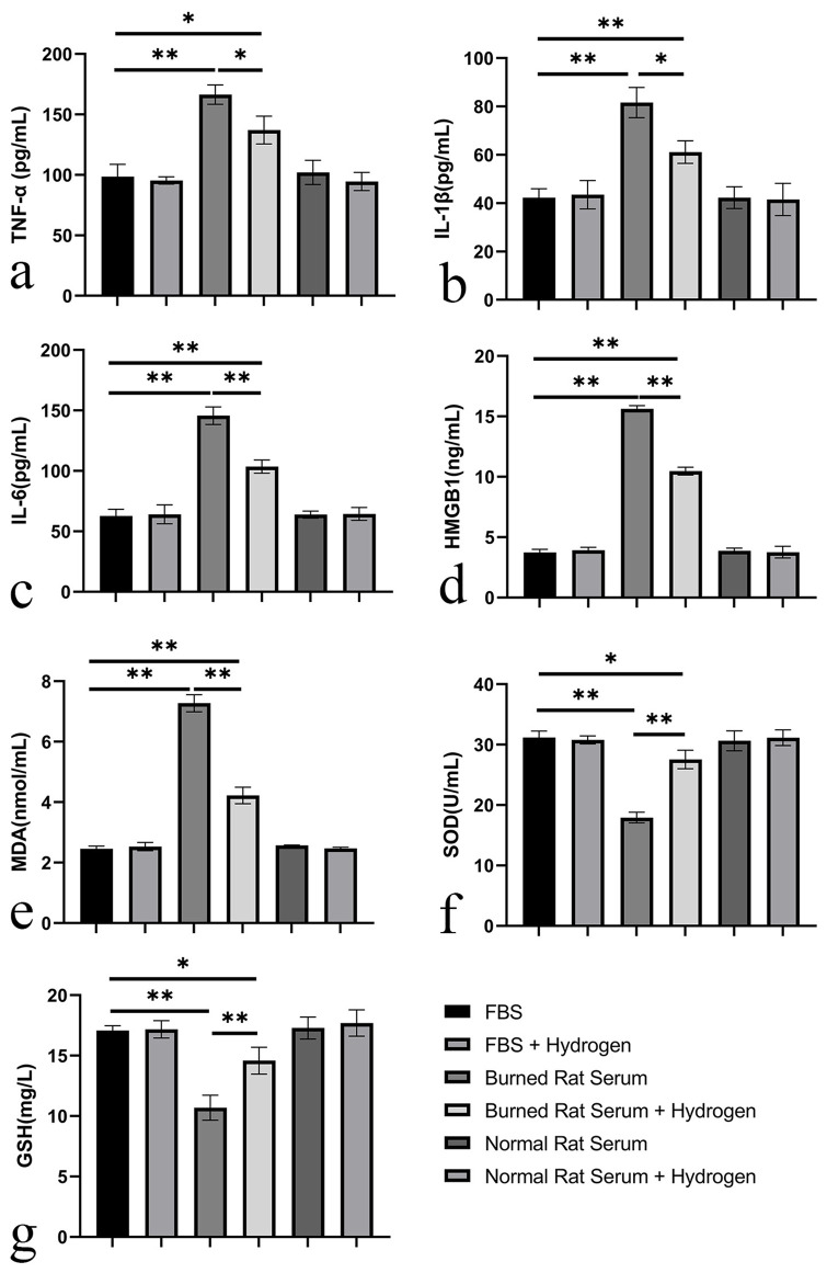
Results: In vitro assays showed that burn serum reduced fibroblast proliferation and increased apoptosis (P < 0.01), which hydrogen gas mitigated by rescuing cell viability and reducing apoptosis (P < 0.01). Enzyme-linked immunosorbent assay revealed burn serum-induced increases in the levels of inflammatory cytokines and oxidative stress markers, with decreases in antioxidant enzymes (P < 0.01), which hydrogen gas reversed (P < 0.05). WB analysis suggested hydrogen gas's anti-inflammatory and proliferative effects by modulating signaling pathways (P < 0.01). In vivo, hydrogen gas treatment considerably improved wound healing, with accelerated contraction and enhanced collagen deposition. Plasma and skin tissue analyses indicated systemic and local anti-inflammatory and antioxidant effects from hydrogen gas.
Conclusion: Hydrogen gas treatment demonstrates potential therapeutic efficacy in the management of postburn sepsis by modulating inflammatory responses, reducing oxidative stress, and promoting wound healing. These findings provide scientific evidence supporting hydrogen gas as an adjunctive treatment strategy for postburn sepsis.
期刊介绍:
The CytoJournal is an open-access peer-reviewed journal committed to publishing high-quality articles in the field of Diagnostic Cytopathology including Molecular aspects. The journal is owned by the Cytopathology Foundation and published by the Scientific Scholar.
 求助内容:
求助内容: 应助结果提醒方式:


