Anna Palomares, Yvonne Espada, Mauricio Tobón Restrepo, Sonia González-Rellán, Rosa Novellas
{"title":"Ultrasonographic Appearance of the Deferent Ducts in Dogs without Genitourinary Disease.","authors":"Anna Palomares, Yvonne Espada, Mauricio Tobón Restrepo, Sonia González-Rellán, Rosa Novellas","doi":"10.1111/vru.70055","DOIUrl":null,"url":null,"abstract":"<p><p>In veterinary medicine, deferent ducts are described as being visible on ultrasound only when pathologically enlarged. Abnormal ultrasonographic enlargement of the deferent ducts has primarily been described secondary to infectious and neoplastic diseases; however, no studies have evaluated the normal ultrasonographic features of these structures. This prospective observational study aimed to describe the ultrasonographic appearance of deferent ducts and assess ultrasound reliability in their identification in intact and neutered dogs without genitourinary disease. The study aimed also to compare ultrasonographic measurements with postmortem anatomical ones and to investigate the relationship between duct identification, size, and intrinsic factors of the patient. The study consisted of ex vivo and in vivo phases in which ultrasonographic assessments of the ducts were conducted at the level of the prostate in longitudinal (location 1) and transverse (location 2) planes and the inguinal canal (location 3) and scrotal region (location 4) in longitudinal planes. A total of 80 deferent ducts were included. The ducts were visible as paired tubular hypoechoic structures delimited by two thin hyperechoic lines, with a target-like appearance on the transverse plane. The deferent ducts were identified in 97.5% of our population in at least one location, with locations 1 and 2 being the most reliable ones. Identification of the ducts was independent of reproductive status; however, reproductive status appeared to be the most significant factor influencing deferent duct size, with neutered dogs exhibiting smaller deferent ducts.</p>","PeriodicalId":23581,"journal":{"name":"Veterinary Radiology & Ultrasound","volume":"66 4","pages":"e70055"},"PeriodicalIF":1.5000,"publicationDate":"2025-07-01","publicationTypes":"Journal Article","fieldsOfStudy":null,"isOpenAccess":false,"openAccessPdf":"https://www.ncbi.nlm.nih.gov/pmc/articles/PMC12137774/pdf/","citationCount":"0","resultStr":null,"platform":"Semanticscholar","paperid":null,"PeriodicalName":"Veterinary Radiology & Ultrasound","FirstCategoryId":"97","ListUrlMain":"https://doi.org/10.1111/vru.70055","RegionNum":2,"RegionCategory":"农林科学","ArticlePicture":[],"TitleCN":null,"AbstractTextCN":null,"PMCID":null,"EPubDate":"","PubModel":"","JCR":"Q2","JCRName":"VETERINARY SCIENCES","Score":null,"Total":0}
引用次数: 0
Abstract
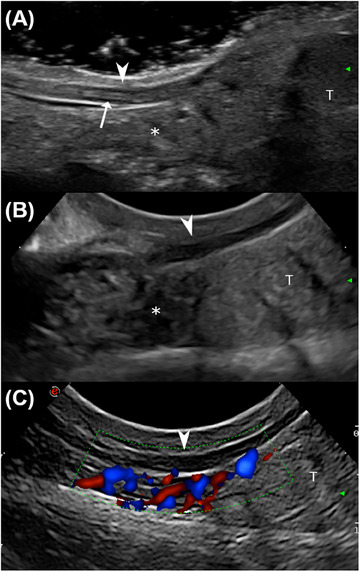
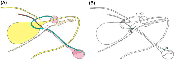
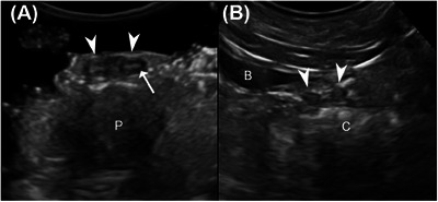
In veterinary medicine, deferent ducts are described as being visible on ultrasound only when pathologically enlarged. Abnormal ultrasonographic enlargement of the deferent ducts has primarily been described secondary to infectious and neoplastic diseases; however, no studies have evaluated the normal ultrasonographic features of these structures. This prospective observational study aimed to describe the ultrasonographic appearance of deferent ducts and assess ultrasound reliability in their identification in intact and neutered dogs without genitourinary disease. The study aimed also to compare ultrasonographic measurements with postmortem anatomical ones and to investigate the relationship between duct identification, size, and intrinsic factors of the patient. The study consisted of ex vivo and in vivo phases in which ultrasonographic assessments of the ducts were conducted at the level of the prostate in longitudinal (location 1) and transverse (location 2) planes and the inguinal canal (location 3) and scrotal region (location 4) in longitudinal planes. A total of 80 deferent ducts were included. The ducts were visible as paired tubular hypoechoic structures delimited by two thin hyperechoic lines, with a target-like appearance on the transverse plane. The deferent ducts were identified in 97.5% of our population in at least one location, with locations 1 and 2 being the most reliable ones. Identification of the ducts was independent of reproductive status; however, reproductive status appeared to be the most significant factor influencing deferent duct size, with neutered dogs exhibiting smaller deferent ducts.
期刊介绍:
Veterinary Radiology & Ultrasound is a bimonthly, international, peer-reviewed, research journal devoted to the fields of veterinary diagnostic imaging and radiation oncology. Established in 1958, it is owned by the American College of Veterinary Radiology and is also the official journal for six affiliate veterinary organizations. Veterinary Radiology & Ultrasound is represented on the International Committee of Medical Journal Editors, World Association of Medical Editors, and Committee on Publication Ethics.
The mission of Veterinary Radiology & Ultrasound is to serve as a leading resource for high quality articles that advance scientific knowledge and standards of clinical practice in the areas of veterinary diagnostic radiology, computed tomography, magnetic resonance imaging, ultrasonography, nuclear imaging, radiation oncology, and interventional radiology. Manuscript types include original investigations, imaging diagnosis reports, review articles, editorials and letters to the Editor. Acceptance criteria include originality, significance, quality, reader interest, composition and adherence to author guidelines.

 求助内容:
求助内容: 应助结果提醒方式:


