{"title":"Low anterior chamber volume as a risk factor in non-arteritic anterior ischemic optic neuropathy.","authors":"Durgul Acan, Beyza Betul Cakar, Eyyup Karahan","doi":"10.3389/fopht.2025.1554279","DOIUrl":null,"url":null,"abstract":"<p><strong>Purpose: </strong>This study aimed to compare the anterior chamber depth (ACD), anterior chamber volume (ACV), and iridocorneal angle (ICA) of the eyes of patients with non-arteritic anterior ischemic optic neuropathy (NAION) and normal eyes.</p><p><strong>Methods: </strong>In this cross-sectional study, 28 patients with NAION who were admitted to our institution were examined. Central corneal thickness (CCT), ACV, ACD, and ICA of all eyes were measured using corneal topography (Sirius, CSO, Italy). Axial lengths (ALs) were measured using an IOL-Master 500 (Carl Zeiss, Meditec). The eyes of these patients were compared with the eyes of 29 healthy individuals of similar age and gender.</p><p><strong>Results: </strong>The mean ALs of the eyes with NAION and those in the control group were not statistically different, measuring 22.95 ± 0.68 mm and 23.13 ± 0.80mm, respectively (p=0.651). While the average ACV was 137.93 ± 41.01 mm<sup>3</sup> in the control group, it was significantly lower at 117.86 ± 22.23 mm<sup>3</sup> in the patients with NAION (p=0.038). The mean ACD, ICA, and CCT values in the control and study groups were not statistically different, with 2.82 ± 0.57 mm and 2.64 ± 0.31 mm, 41.62 ± 6.99° and 40.14 ± 7.04°, and 542.48 ± 19.39µm and 544.68 ± 31.26 µm, respectively (p1 = 0.236, p2 = 0.693, and p3 = 0.959). No statistical differences were found between the eyes with NAION and their fellow eyes in terms of AL, CCT, ACD, ACV, and ICA (p>0.05).</p><p><strong>Conclusion: </strong>Differences in anterior segment morphology were observed in eyes with NAION compared to healthy eyes. Decreased ACV may be a risk factor for NAION.</p>","PeriodicalId":73096,"journal":{"name":"Frontiers in ophthalmology","volume":"5 ","pages":"1554279"},"PeriodicalIF":0.9000,"publicationDate":"2025-05-20","publicationTypes":"Journal Article","fieldsOfStudy":null,"isOpenAccess":false,"openAccessPdf":"https://www.ncbi.nlm.nih.gov/pmc/articles/PMC12129766/pdf/","citationCount":"0","resultStr":null,"platform":"Semanticscholar","paperid":null,"PeriodicalName":"Frontiers in ophthalmology","FirstCategoryId":"1085","ListUrlMain":"https://doi.org/10.3389/fopht.2025.1554279","RegionNum":0,"RegionCategory":null,"ArticlePicture":[],"TitleCN":null,"AbstractTextCN":null,"PMCID":null,"EPubDate":"2025/1/1 0:00:00","PubModel":"eCollection","JCR":"","JCRName":"","Score":null,"Total":0}
引用次数: 0
Abstract
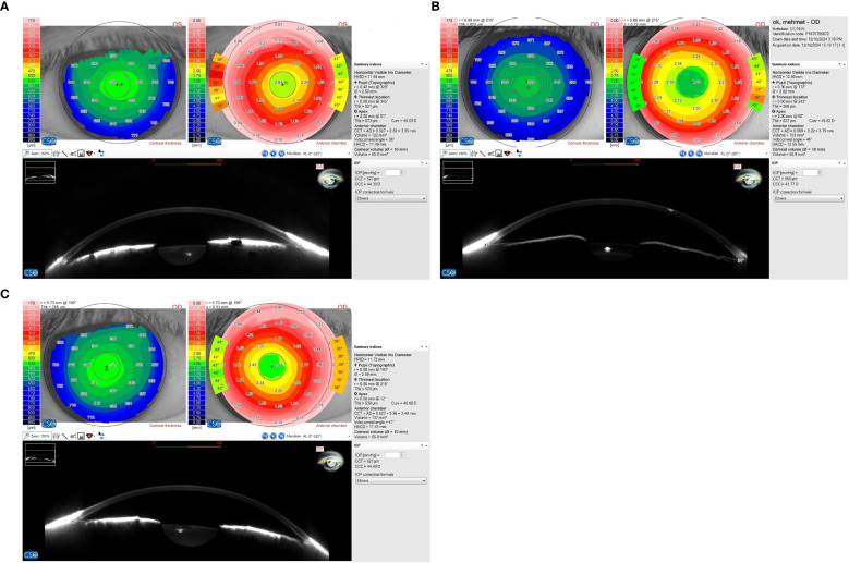
Purpose: This study aimed to compare the anterior chamber depth (ACD), anterior chamber volume (ACV), and iridocorneal angle (ICA) of the eyes of patients with non-arteritic anterior ischemic optic neuropathy (NAION) and normal eyes.
Methods: In this cross-sectional study, 28 patients with NAION who were admitted to our institution were examined. Central corneal thickness (CCT), ACV, ACD, and ICA of all eyes were measured using corneal topography (Sirius, CSO, Italy). Axial lengths (ALs) were measured using an IOL-Master 500 (Carl Zeiss, Meditec). The eyes of these patients were compared with the eyes of 29 healthy individuals of similar age and gender.
Results: The mean ALs of the eyes with NAION and those in the control group were not statistically different, measuring 22.95 ± 0.68 mm and 23.13 ± 0.80mm, respectively (p=0.651). While the average ACV was 137.93 ± 41.01 mm3 in the control group, it was significantly lower at 117.86 ± 22.23 mm3 in the patients with NAION (p=0.038). The mean ACD, ICA, and CCT values in the control and study groups were not statistically different, with 2.82 ± 0.57 mm and 2.64 ± 0.31 mm, 41.62 ± 6.99° and 40.14 ± 7.04°, and 542.48 ± 19.39µm and 544.68 ± 31.26 µm, respectively (p1 = 0.236, p2 = 0.693, and p3 = 0.959). No statistical differences were found between the eyes with NAION and their fellow eyes in terms of AL, CCT, ACD, ACV, and ICA (p>0.05).
Conclusion: Differences in anterior segment morphology were observed in eyes with NAION compared to healthy eyes. Decreased ACV may be a risk factor for NAION.

 求助内容:
求助内容: 应助结果提醒方式:


