Evaluation of lymph node metastasis in cervical cancer: A retrospective comparison of preoperative MRI and PET/CT with postoperative histopathology results.
Süleyman Özen, Ergul Demircivi, Abdulkadir Turgut, Muzaffer Sancı
{"title":"Evaluation of lymph node metastasis in cervical cancer: A retrospective comparison of preoperative MRI and PET/CT with postoperative histopathology results.","authors":"Süleyman Özen, Ergul Demircivi, Abdulkadir Turgut, Muzaffer Sancı","doi":"10.4274/tjod.galenos.2025.27482","DOIUrl":null,"url":null,"abstract":"<p><strong>Objective: </strong>The aim of this study is to assess the diagnostic performance of positron emission tomography/computed tomography (PET/CT) and magnetic resonance imaging (MRI) in detecting pelvic and paraaortic lymph node involvement in cervical cancer patients by correlating imaging results with surgical pathology findings.</p><p><strong>Materials and methods: </strong>A retrospective analysis was conducted on cervical cancer patients treated at İstanbul Medeniyet University Prof. Dr. Süleyman Yalçın City Hospital from 2016 to 2022. Patients who underwent preoperative PET/CT or MRI imaging and subsequent lymph node dissection were included. Sensitivity, specificity, positive predictive value, and negative predictive value were calculated for each imaging modality.</p><p><strong>Results: </strong>Of the 75 cases reviewed, 52 met the inclusion criteria. PET/CT had higher specificity (94.1%) than MRI (82.4%), while MRI demonstrated greater sensitivity (55.6% vs. 50%). False-negative rates were 15.3% for MRI and 17.3% for PET/CT. Receiver operating characteristic analysis indicated an area under the curve of 0.78 for PET/CT and 0.69 for MRI. No statistically significant differences in sensitivity or specificity were observed, with both modalities showing complementary strengths.</p><p><strong>Conclusion: </strong>MRI and PET/CT each contribute significantly to preoperative cervical cancer evaluation, with MRI favored for local assessment and PET/CT for nodal detection. Combining both modalities enhances diagnostic accuracy. Further prospective research is required to confirm and strengthen these results. and improve imaging strategies for clinical practice.</p>","PeriodicalId":45340,"journal":{"name":"Turkish Journal of Obstetrics and Gynecology","volume":"22 2","pages":"129-133"},"PeriodicalIF":1.3000,"publicationDate":"2025-06-04","publicationTypes":"Journal Article","fieldsOfStudy":null,"isOpenAccess":false,"openAccessPdf":"https://www.ncbi.nlm.nih.gov/pmc/articles/PMC12136126/pdf/","citationCount":"0","resultStr":null,"platform":"Semanticscholar","paperid":null,"PeriodicalName":"Turkish Journal of Obstetrics and Gynecology","FirstCategoryId":"1085","ListUrlMain":"https://doi.org/10.4274/tjod.galenos.2025.27482","RegionNum":0,"RegionCategory":null,"ArticlePicture":[],"TitleCN":null,"AbstractTextCN":null,"PMCID":null,"EPubDate":"","PubModel":"","JCR":"Q4","JCRName":"OBSTETRICS & GYNECOLOGY","Score":null,"Total":0}
引用次数: 0
Abstract
Objective: The aim of this study is to assess the diagnostic performance of positron emission tomography/computed tomography (PET/CT) and magnetic resonance imaging (MRI) in detecting pelvic and paraaortic lymph node involvement in cervical cancer patients by correlating imaging results with surgical pathology findings.
Materials and methods: A retrospective analysis was conducted on cervical cancer patients treated at İstanbul Medeniyet University Prof. Dr. Süleyman Yalçın City Hospital from 2016 to 2022. Patients who underwent preoperative PET/CT or MRI imaging and subsequent lymph node dissection were included. Sensitivity, specificity, positive predictive value, and negative predictive value were calculated for each imaging modality.
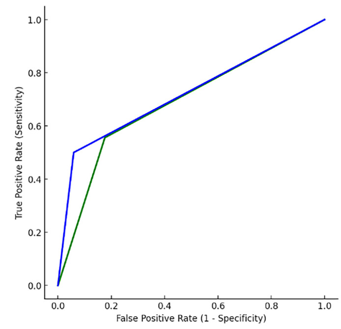
Results: Of the 75 cases reviewed, 52 met the inclusion criteria. PET/CT had higher specificity (94.1%) than MRI (82.4%), while MRI demonstrated greater sensitivity (55.6% vs. 50%). False-negative rates were 15.3% for MRI and 17.3% for PET/CT. Receiver operating characteristic analysis indicated an area under the curve of 0.78 for PET/CT and 0.69 for MRI. No statistically significant differences in sensitivity or specificity were observed, with both modalities showing complementary strengths.
Conclusion: MRI and PET/CT each contribute significantly to preoperative cervical cancer evaluation, with MRI favored for local assessment and PET/CT for nodal detection. Combining both modalities enhances diagnostic accuracy. Further prospective research is required to confirm and strengthen these results. and improve imaging strategies for clinical practice.

 求助内容:
求助内容: 应助结果提醒方式:


