Wout J Claassen, Isabel M van Ruijven, Marloes van den Berg, Rianne J Baelde, Alexcia Fortes Monteiro, Rajvi M N Balesar, Sylvia W Hania, Donald L van der Peet, Peter J M Weijs, Coen A C Ottenheijm, Sandra N Stapel
{"title":"In vitro and in vivo muscle mass and strength during the first week of critical illness.","authors":"Wout J Claassen, Isabel M van Ruijven, Marloes van den Berg, Rianne J Baelde, Alexcia Fortes Monteiro, Rajvi M N Balesar, Sylvia W Hania, Donald L van der Peet, Peter J M Weijs, Coen A C Ottenheijm, Sandra N Stapel","doi":"10.1186/s40635-025-00755-7","DOIUrl":null,"url":null,"abstract":"<p><strong>Background: </strong>Loss of muscle mass and strength is provoked by critical illness. Our primary aim was to study the development of muscle atrophy and weakness in vitro in isolated myofibers and in vivo muscle mass and in vitro muscle strength during the first week of critical illness. Furthermore, we explored how in vitro muscle strength compares to healthy controls. Finally, we studied correlations between in vitro muscle mass and strength and in vivo muscle mass in critically ill patients.</p><p><strong>Methods: </strong>We performed a secondary analysis using data from a randomized controlled trial. We studied contractile force of single myofibers isolated from muscle biopsies around admission (day 1-3) and around 1 week after inclusion (day 8-10). Furthermore, we studied myofiber cross-sectional area (CSA), proportion of fast-twitch myofibers, bio-electrical impedance analysis-derived fat-free mass index (FFMI), ultrasound-derived quadriceps muscle layer thickness (QMLT) and diaphragm thickness. In the control group, only contractile force outcomes were available.</p><p><strong>Results: </strong>In total, ten ICU patients had two muscle biopsies taken. Maximum force of both fast and slow-twitch myofibers was reduced at day 8-10 compared to day 1-3, even though there were no differences in normalized force and calcium sensitivity. FFM and QMLT did not change over time, nor were there differences between groups. Compared to healthy controls, maximum force of myofibers was lower in the ICU group at day 8-10 in both slow and fast-twitch myofibers, while the calcium sensitivity of force was lower in slow-twitch myofibers. We found a significant correlation between myofiber CSA vs. FFMI (r = 0.68) and maximum force of the fast-twitch fibers vs. QMLT (r = 0.72).</p><p><strong>Conclusions: </strong>During the first week of critical illness, maximum force declined over time, while no other in vitro parameters changed. We found a moderate correlation between myofiber CSA vs. FFMI and maximum force of the fast-twitch fibers vs. QMLT.</p>","PeriodicalId":13750,"journal":{"name":"Intensive Care Medicine Experimental","volume":"13 1","pages":"57"},"PeriodicalIF":2.8000,"publicationDate":"2025-06-03","publicationTypes":"Journal Article","fieldsOfStudy":null,"isOpenAccess":false,"openAccessPdf":"https://www.ncbi.nlm.nih.gov/pmc/articles/PMC12133653/pdf/","citationCount":"0","resultStr":null,"platform":"Semanticscholar","paperid":null,"PeriodicalName":"Intensive Care Medicine Experimental","FirstCategoryId":"1085","ListUrlMain":"https://doi.org/10.1186/s40635-025-00755-7","RegionNum":0,"RegionCategory":null,"ArticlePicture":[],"TitleCN":null,"AbstractTextCN":null,"PMCID":null,"EPubDate":"","PubModel":"","JCR":"Q2","JCRName":"CRITICAL CARE MEDICINE","Score":null,"Total":0}
引用次数: 0
Abstract
Background: Loss of muscle mass and strength is provoked by critical illness. Our primary aim was to study the development of muscle atrophy and weakness in vitro in isolated myofibers and in vivo muscle mass and in vitro muscle strength during the first week of critical illness. Furthermore, we explored how in vitro muscle strength compares to healthy controls. Finally, we studied correlations between in vitro muscle mass and strength and in vivo muscle mass in critically ill patients.
Methods: We performed a secondary analysis using data from a randomized controlled trial. We studied contractile force of single myofibers isolated from muscle biopsies around admission (day 1-3) and around 1 week after inclusion (day 8-10). Furthermore, we studied myofiber cross-sectional area (CSA), proportion of fast-twitch myofibers, bio-electrical impedance analysis-derived fat-free mass index (FFMI), ultrasound-derived quadriceps muscle layer thickness (QMLT) and diaphragm thickness. In the control group, only contractile force outcomes were available.
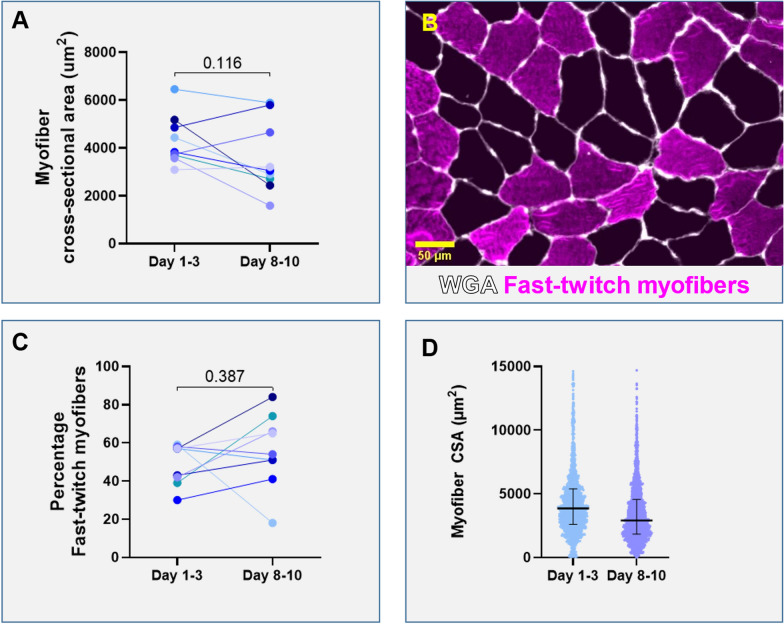
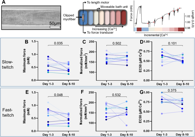
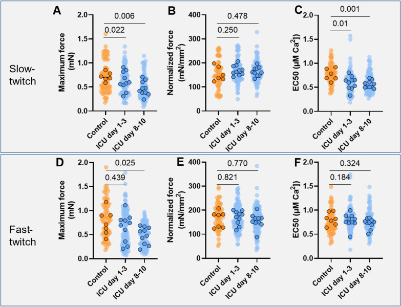
Results: In total, ten ICU patients had two muscle biopsies taken. Maximum force of both fast and slow-twitch myofibers was reduced at day 8-10 compared to day 1-3, even though there were no differences in normalized force and calcium sensitivity. FFM and QMLT did not change over time, nor were there differences between groups. Compared to healthy controls, maximum force of myofibers was lower in the ICU group at day 8-10 in both slow and fast-twitch myofibers, while the calcium sensitivity of force was lower in slow-twitch myofibers. We found a significant correlation between myofiber CSA vs. FFMI (r = 0.68) and maximum force of the fast-twitch fibers vs. QMLT (r = 0.72).
Conclusions: During the first week of critical illness, maximum force declined over time, while no other in vitro parameters changed. We found a moderate correlation between myofiber CSA vs. FFMI and maximum force of the fast-twitch fibers vs. QMLT.
 求助内容:
求助内容: 应助结果提醒方式:


