Heterochronic pelvic high-grade myxoinflammatory fibroblastic sarcoma and uterine endometroid carcinoma harboring common gene mutations: a rare case report with genomic analysis.
{"title":"Heterochronic pelvic high-grade myxoinflammatory fibroblastic sarcoma and uterine endometroid carcinoma harboring common gene mutations: a rare case report with genomic analysis.","authors":"Yuriko Higashi, Mika Mizuno, Ikumi Kitazono, Toshiaki Akahane, Takashi Tasaki, Hirotsugu Noguchi, Masanori Hisaoka, Hiroaki Kobayashi, Akihide Tanimoto","doi":"10.1186/s13000-025-01669-4","DOIUrl":null,"url":null,"abstract":"<p><strong>Objective: </strong>This report presents a rare case involving an extreme epithelial-to-mesenchymal transition, in which a specific type of sarcoma developed heterochronically as a recurrence of endometrioid carcinoma.</p><p><strong>Case presentation: </strong>A female in her 50's presented with abnormal genital bleeding, and an endometrial biopsy revealed endometrioid carcinoma. Following the diagnosis of stage IA endometrioid carcinoma according to the 2008 classification system of the International Federation of Gynecology and Obstetrics, a robot-assisted simple hysterectomy, bilateral salpingo-oophorectomy, and sentinel lymph node navigation surgery were performed. Six months postoperatively, a tumor mass developed in the pelvis. A transrectal needle biopsy revealed spindle cell proliferation, and pelvic tumor resection was conducted for diagnostic therapy. The patient received no adjuvant chemotherapy or radiotherapy after the second surgery and remained free of tumor recurrence for 8 months. The resected yellowish solid tumor mass, measuring 16 × 12 × 9 cm, exhibited hemorrhage, necrosis, and cystic degeneration and was composed of fascicular proliferation of spindle tumor cells showing nuclear pleomorphism and frequent mitotic figures within a myxoid and inflammatory stroma. No epithelial component or organoid patterns were observed. Immunohistochemically, the tumor cells were positive for factor XIIIa, CD10, and cyclin D1, but negative for keratins (AE1/AE3 and CAM5.2) and other specific markers, supporting a diagnosis of high-grade myxoinflammatory fibroblastic sarcoma (MIFS).</p><p><strong>Conclusion: </strong>Genomic analysis revealed identical mutations in PTEN, PIK3R1, CDKN2 A, and TP53 in both the primary uterine endometrioid carcinoma and heterochronic pelvic MIFS. An integrative approach involving histology, immunohistochemistry, and genomic analysis is critical for elucidating the pathogenesis of rare pelvic and uterine tumors.</p>","PeriodicalId":11237,"journal":{"name":"Diagnostic Pathology","volume":"20 1","pages":"71"},"PeriodicalIF":2.3000,"publicationDate":"2025-06-03","publicationTypes":"Journal Article","fieldsOfStudy":null,"isOpenAccess":false,"openAccessPdf":"https://www.ncbi.nlm.nih.gov/pmc/articles/PMC12131376/pdf/","citationCount":"0","resultStr":null,"platform":"Semanticscholar","paperid":null,"PeriodicalName":"Diagnostic Pathology","FirstCategoryId":"3","ListUrlMain":"https://doi.org/10.1186/s13000-025-01669-4","RegionNum":3,"RegionCategory":"医学","ArticlePicture":[],"TitleCN":null,"AbstractTextCN":null,"PMCID":null,"EPubDate":"","PubModel":"","JCR":"Q2","JCRName":"PATHOLOGY","Score":null,"Total":0}
引用次数: 0
Abstract
Objective: This report presents a rare case involving an extreme epithelial-to-mesenchymal transition, in which a specific type of sarcoma developed heterochronically as a recurrence of endometrioid carcinoma.
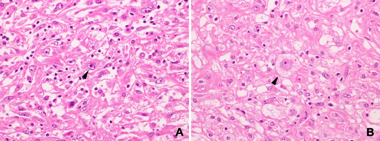
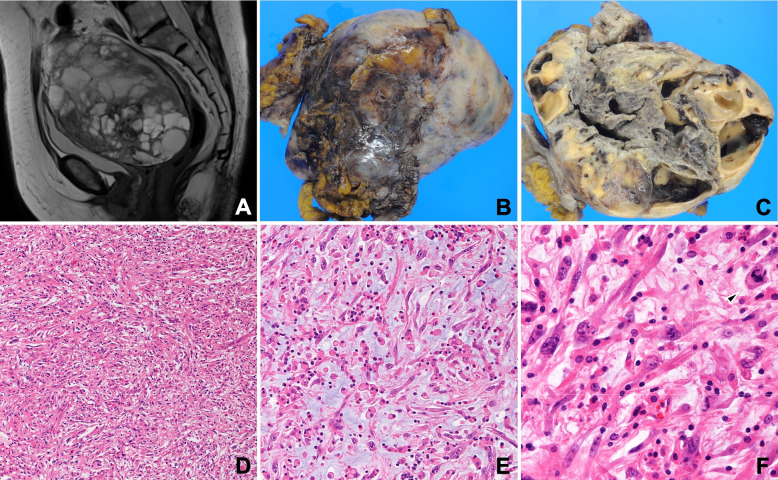
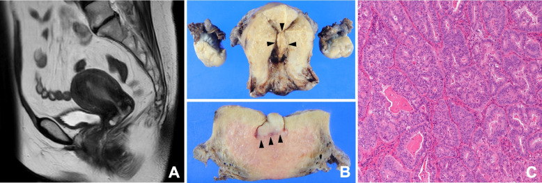
Case presentation: A female in her 50's presented with abnormal genital bleeding, and an endometrial biopsy revealed endometrioid carcinoma. Following the diagnosis of stage IA endometrioid carcinoma according to the 2008 classification system of the International Federation of Gynecology and Obstetrics, a robot-assisted simple hysterectomy, bilateral salpingo-oophorectomy, and sentinel lymph node navigation surgery were performed. Six months postoperatively, a tumor mass developed in the pelvis. A transrectal needle biopsy revealed spindle cell proliferation, and pelvic tumor resection was conducted for diagnostic therapy. The patient received no adjuvant chemotherapy or radiotherapy after the second surgery and remained free of tumor recurrence for 8 months. The resected yellowish solid tumor mass, measuring 16 × 12 × 9 cm, exhibited hemorrhage, necrosis, and cystic degeneration and was composed of fascicular proliferation of spindle tumor cells showing nuclear pleomorphism and frequent mitotic figures within a myxoid and inflammatory stroma. No epithelial component or organoid patterns were observed. Immunohistochemically, the tumor cells were positive for factor XIIIa, CD10, and cyclin D1, but negative for keratins (AE1/AE3 and CAM5.2) and other specific markers, supporting a diagnosis of high-grade myxoinflammatory fibroblastic sarcoma (MIFS).
Conclusion: Genomic analysis revealed identical mutations in PTEN, PIK3R1, CDKN2 A, and TP53 in both the primary uterine endometrioid carcinoma and heterochronic pelvic MIFS. An integrative approach involving histology, immunohistochemistry, and genomic analysis is critical for elucidating the pathogenesis of rare pelvic and uterine tumors.
期刊介绍:
Diagnostic Pathology is an open access, peer-reviewed, online journal that considers research in surgical and clinical pathology, immunology, and biology, with a special focus on cutting-edge approaches in diagnostic pathology and tissue-based therapy. The journal covers all aspects of surgical pathology, including classic diagnostic pathology, prognosis-related diagnosis (tumor stages, prognosis markers, such as MIB-percentage, hormone receptors, etc.), and therapy-related findings. The journal also focuses on the technological aspects of pathology, including molecular biology techniques, morphometry aspects (stereology, DNA analysis, syntactic structure analysis), communication aspects (telecommunication, virtual microscopy, virtual pathology institutions, etc.), and electronic education and quality assurance (for example interactive publication, on-line references with automated updating, etc.).

 求助内容:
求助内容: 应助结果提醒方式:


