Andrea Sturchio, Jonas E Svensson, Mikael Tiger, Anton Forsberg Morén, Andrea Varrone, Per Svenningsson, Yoshiro Okubo, Amane Tateno
{"title":"[<sup>18</sup>F]FE-PE2I PET is a diagnostic tool in dementia with Lewy bodies.","authors":"Andrea Sturchio, Jonas E Svensson, Mikael Tiger, Anton Forsberg Morén, Andrea Varrone, Per Svenningsson, Yoshiro Okubo, Amane Tateno","doi":"10.1002/pcn5.70123","DOIUrl":null,"url":null,"abstract":"<p><strong>Aim: </strong>Dementia with Lewy bodies (DLB) is characterized by motor and non-motor symptoms. The degeneration of the dopaminergic pathway is a hallmark of DLB; for this reason, we aimed to study a recent dopamine transporter (DAT) positron emission tomography (PET) radioligand as a diagnostic tool for DLB.</p><p><strong>Methods: </strong>In this study, we used DAT-PET with the radioligand [<sup>18</sup>F]FE-PE2I to distinguish DLB subjects from healthy controls (HCs). We also aimed to analyze how DAT binding correlated with clinical features, amyloid load, measured by PET, and cardiac metaiodobenzylguanidine scintigraphy (MIBG).</p><p><strong>Results: </strong>Binding potential (<i>BP</i> <sub>ND</sub>) values of [<sup>18</sup>F]FE-PE2I were higher in HCs versus DLB in striatum (1.82 ± 0.34 vs. 1.15 ± 0.34; <i>p</i> < 0.001; 95% Confidence Interval [CI]: 0.40-0.96), putamen (2.2 ± 0.36 vs. 1.41 ± 0.51; <i>p</i> < 0.001; 95% CI: 0.39-1.17), caudate (1.38 ± 0.30 vs. 0.88 ± 0.20; <i>p</i> < 0.001; 95% CI: 0.28-0.70), and substantia nigra (0.49 ± 0.091 vs. 0.42 ± 0.084; <i>p</i> = 0.0437; 95% CI: 0.003 to 0.14). After adjusting for age, substantia nigra did not differ between DLB and HCs (<i>p</i>: 0.46; 95% CI: -0.049 to 0.11); however, <i>BP</i> <sub>ND</sub> values between DLB and HC in striatum (<i>p</i>: <0.001; 95% CI: 0.25-0.85), putamen (<i>p</i>: 0.0012; 95% CI: 0.31-1.13), and caudate (<i>p</i>: 0.0027; 95% CI: 0.13-0.55) were still significant. Striatum was the best area to correctly classify DLB subjects versus HC compared to the putamen, caudate, and substantia nigra (area under the curve = 0.95, 0.90, 0.93, and 0.73, respectively; 95 CI: 0.87-1.00, 0.79-1.00, 0.84-1.00, 0.55-0.92, respectively). Subjects with altered MIBG showed lower <i>BP</i> <sub>ND</sub> compared to subjects with normal MIBG in the putamen.</p><p><strong>Conclusion: </strong>Our study showed that [<sup>18</sup>F]FE-PE2I PET represents a potential diagnostic tool with high accuracy in discriminating DLB patients versus HC, which is valuable for clinical practice.</p>","PeriodicalId":74405,"journal":{"name":"PCN reports : psychiatry and clinical neurosciences","volume":"4 2","pages":"e70123"},"PeriodicalIF":0.9000,"publicationDate":"2025-06-02","publicationTypes":"Journal Article","fieldsOfStudy":null,"isOpenAccess":false,"openAccessPdf":"https://www.ncbi.nlm.nih.gov/pmc/articles/PMC12128163/pdf/","citationCount":"0","resultStr":null,"platform":"Semanticscholar","paperid":null,"PeriodicalName":"PCN reports : psychiatry and clinical neurosciences","FirstCategoryId":"1085","ListUrlMain":"https://doi.org/10.1002/pcn5.70123","RegionNum":0,"RegionCategory":null,"ArticlePicture":[],"TitleCN":null,"AbstractTextCN":null,"PMCID":null,"EPubDate":"2025/6/1 0:00:00","PubModel":"eCollection","JCR":"","JCRName":"","Score":null,"Total":0}
引用次数: 0
Abstract
Aim: Dementia with Lewy bodies (DLB) is characterized by motor and non-motor symptoms. The degeneration of the dopaminergic pathway is a hallmark of DLB; for this reason, we aimed to study a recent dopamine transporter (DAT) positron emission tomography (PET) radioligand as a diagnostic tool for DLB.
Methods: In this study, we used DAT-PET with the radioligand [18F]FE-PE2I to distinguish DLB subjects from healthy controls (HCs). We also aimed to analyze how DAT binding correlated with clinical features, amyloid load, measured by PET, and cardiac metaiodobenzylguanidine scintigraphy (MIBG).
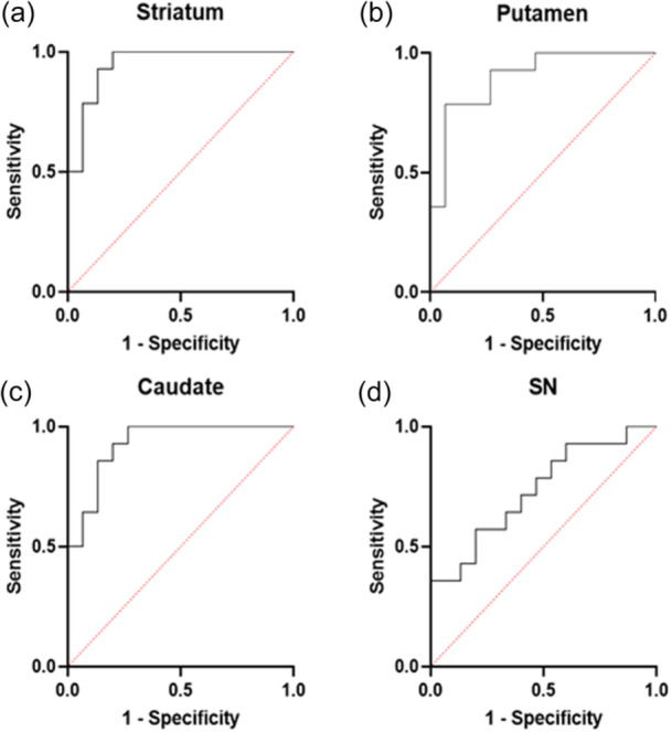
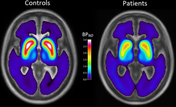
Results: Binding potential (BPND) values of [18F]FE-PE2I were higher in HCs versus DLB in striatum (1.82 ± 0.34 vs. 1.15 ± 0.34; p < 0.001; 95% Confidence Interval [CI]: 0.40-0.96), putamen (2.2 ± 0.36 vs. 1.41 ± 0.51; p < 0.001; 95% CI: 0.39-1.17), caudate (1.38 ± 0.30 vs. 0.88 ± 0.20; p < 0.001; 95% CI: 0.28-0.70), and substantia nigra (0.49 ± 0.091 vs. 0.42 ± 0.084; p = 0.0437; 95% CI: 0.003 to 0.14). After adjusting for age, substantia nigra did not differ between DLB and HCs (p: 0.46; 95% CI: -0.049 to 0.11); however, BPND values between DLB and HC in striatum (p: <0.001; 95% CI: 0.25-0.85), putamen (p: 0.0012; 95% CI: 0.31-1.13), and caudate (p: 0.0027; 95% CI: 0.13-0.55) were still significant. Striatum was the best area to correctly classify DLB subjects versus HC compared to the putamen, caudate, and substantia nigra (area under the curve = 0.95, 0.90, 0.93, and 0.73, respectively; 95 CI: 0.87-1.00, 0.79-1.00, 0.84-1.00, 0.55-0.92, respectively). Subjects with altered MIBG showed lower BPND compared to subjects with normal MIBG in the putamen.
Conclusion: Our study showed that [18F]FE-PE2I PET represents a potential diagnostic tool with high accuracy in discriminating DLB patients versus HC, which is valuable for clinical practice.


 求助内容:
求助内容: 应助结果提醒方式:


