{"title":"The Poor Prognostic Stigma of Hepatic Superscan on <sup>18</sup>F-FDG PET Imaging.","authors":"Dhuha Al-Adhami, Malik Juweid, Akram Al-Ibraheem","doi":"10.4274/mirt.galenos.2024.56689","DOIUrl":null,"url":null,"abstract":"<p><p>Pancreatic exocrine carcinoma (PEC) is a lethal malignancy with high mortality rates because of its aggressive nature and frequent late-stage diagnosis. When histopathological diagnosis becomes unfeasible because of patient deterioration, clinicians must rely on clinical, biochemical, and radiological findings. This case report describes a 78-year-old woman with aggressive PEC visualized through <sup>18</sup>F-fluorodeoxyglucose (<sup>18</sup>F-FDG) positron emission tomography/computed tomography. The imaging revealed an intensely hypermetabolic head mass, hepatic superscan, and hypermetabolic abdominal lymphadenopathy. Despite strong clinical indicators suggesting stage IV PEC, rapid disease progression and patient demise precluded histopathological confirmation, emphasizing the poor prognosis associated with hepatic superscan in this context.</p>","PeriodicalId":44681,"journal":{"name":"Molecular Imaging and Radionuclide Therapy","volume":"34 2","pages":"149-151"},"PeriodicalIF":1.1000,"publicationDate":"2025-06-03","publicationTypes":"Journal Article","fieldsOfStudy":null,"isOpenAccess":false,"openAccessPdf":"https://www.ncbi.nlm.nih.gov/pmc/articles/PMC12134963/pdf/","citationCount":"0","resultStr":null,"platform":"Semanticscholar","paperid":null,"PeriodicalName":"Molecular Imaging and Radionuclide Therapy","FirstCategoryId":"1085","ListUrlMain":"https://doi.org/10.4274/mirt.galenos.2024.56689","RegionNum":0,"RegionCategory":null,"ArticlePicture":[],"TitleCN":null,"AbstractTextCN":null,"PMCID":null,"EPubDate":"","PubModel":"","JCR":"Q4","JCRName":"RADIOLOGY, NUCLEAR MEDICINE & MEDICAL IMAGING","Score":null,"Total":0}
引用次数: 0
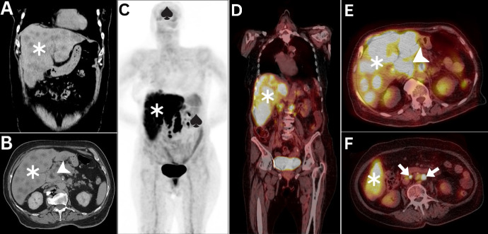
Abstract
Pancreatic exocrine carcinoma (PEC) is a lethal malignancy with high mortality rates because of its aggressive nature and frequent late-stage diagnosis. When histopathological diagnosis becomes unfeasible because of patient deterioration, clinicians must rely on clinical, biochemical, and radiological findings. This case report describes a 78-year-old woman with aggressive PEC visualized through 18F-fluorodeoxyglucose (18F-FDG) positron emission tomography/computed tomography. The imaging revealed an intensely hypermetabolic head mass, hepatic superscan, and hypermetabolic abdominal lymphadenopathy. Despite strong clinical indicators suggesting stage IV PEC, rapid disease progression and patient demise precluded histopathological confirmation, emphasizing the poor prognosis associated with hepatic superscan in this context.
期刊介绍:
Molecular Imaging and Radionuclide Therapy (Mol Imaging Radionucl Ther, MIRT) is publishes original research articles, invited reviews, editorials, short communications, letters, consensus statements, guidelines and case reports with a literature review on the topic, in the field of molecular imaging, multimodality imaging, nuclear medicine, radionuclide therapy, radiopharmacy, medical physics, dosimetry and radiobiology.

 求助内容:
求助内容: 应助结果提醒方式:


