{"title":"Combined <sup>68</sup>Ga-PSMA PET/CT and mpMRI Findings Improve Tumor Localization and Biopsy Guidance in the Initial Diagnosis of Prostate Cancer.","authors":"Yavor Gramatikov, Petia Nikolova, Mihaela Ilcheva, Valentin Yotovski, Nikolay Halachev, Valeria Hadzhiyska","doi":"10.4274/mirt.galenos.2024.75547","DOIUrl":null,"url":null,"abstract":"<p><p>Gallium-68 (<sup>68</sup>Ga)-prostate-specific membrane antigen (PSMA) positron emission tomography/computed tomography (PET/CT) is a relatively new imaging modality that has already proved its role in the initial staging of prostate cancer and in biochemical recurrence following definitive primary therapy. Furthermore, emerging data several ongoing studies demonstrate its potential role in the primary diagnosis of this malignancy. We present a 67-year-old male patient with increasing clinical suspicion of prostate cancer despite a previous negative prostate gland biopsy. He was referred to our nuclear medicine department for a <sup>68</sup>Ga-PSMA PET/CT with the aim of improving tumor localization and assisting in the guidance of repeat prostate biopsy. One month before presentation of elevated prostate-specific antigen levels, he underwent multiparametric magnetic resonance imaging (mpMRI), which revealed a prostate imaging reporting and data system 4 lesion in the right lobe of the prostate gland. The MRI lesion completely matched a PRIMARY score 5 lesion registered by PET/CT. Furthermore, we detected another suspicious finding in the left lobe (PRIMARY score 4). The patient underwent PSMA PET/CT-guided MRI/ultrasonography fusion transperineal biopsy of both lesions. The latter were histologically confirmed as prostate carcinoma with a Gleason score of 3+4 =7.</p>","PeriodicalId":44681,"journal":{"name":"Molecular Imaging and Radionuclide Therapy","volume":"34 2","pages":"146-148"},"PeriodicalIF":1.1000,"publicationDate":"2025-06-03","publicationTypes":"Journal Article","fieldsOfStudy":null,"isOpenAccess":false,"openAccessPdf":"https://www.ncbi.nlm.nih.gov/pmc/articles/PMC12134961/pdf/","citationCount":"0","resultStr":null,"platform":"Semanticscholar","paperid":null,"PeriodicalName":"Molecular Imaging and Radionuclide Therapy","FirstCategoryId":"1085","ListUrlMain":"https://doi.org/10.4274/mirt.galenos.2024.75547","RegionNum":0,"RegionCategory":null,"ArticlePicture":[],"TitleCN":null,"AbstractTextCN":null,"PMCID":null,"EPubDate":"","PubModel":"","JCR":"Q4","JCRName":"RADIOLOGY, NUCLEAR MEDICINE & MEDICAL IMAGING","Score":null,"Total":0}
引用次数: 0
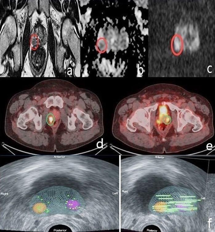
Abstract
Gallium-68 (68Ga)-prostate-specific membrane antigen (PSMA) positron emission tomography/computed tomography (PET/CT) is a relatively new imaging modality that has already proved its role in the initial staging of prostate cancer and in biochemical recurrence following definitive primary therapy. Furthermore, emerging data several ongoing studies demonstrate its potential role in the primary diagnosis of this malignancy. We present a 67-year-old male patient with increasing clinical suspicion of prostate cancer despite a previous negative prostate gland biopsy. He was referred to our nuclear medicine department for a 68Ga-PSMA PET/CT with the aim of improving tumor localization and assisting in the guidance of repeat prostate biopsy. One month before presentation of elevated prostate-specific antigen levels, he underwent multiparametric magnetic resonance imaging (mpMRI), which revealed a prostate imaging reporting and data system 4 lesion in the right lobe of the prostate gland. The MRI lesion completely matched a PRIMARY score 5 lesion registered by PET/CT. Furthermore, we detected another suspicious finding in the left lobe (PRIMARY score 4). The patient underwent PSMA PET/CT-guided MRI/ultrasonography fusion transperineal biopsy of both lesions. The latter were histologically confirmed as prostate carcinoma with a Gleason score of 3+4 =7.
期刊介绍:
Molecular Imaging and Radionuclide Therapy (Mol Imaging Radionucl Ther, MIRT) is publishes original research articles, invited reviews, editorials, short communications, letters, consensus statements, guidelines and case reports with a literature review on the topic, in the field of molecular imaging, multimodality imaging, nuclear medicine, radionuclide therapy, radiopharmacy, medical physics, dosimetry and radiobiology.

 求助内容:
求助内容: 应助结果提醒方式:


