Siven Kar, Harshita Gupta, Nusrat Shaikh, Vikram R Lele
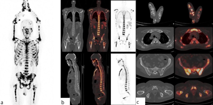
{"title":"<sup>68</sup>Ga-PENTIXAFOR PET/CT Captures Superscan in Refractory Multiple Myeloma.","authors":"Siven Kar, Harshita Gupta, Nusrat Shaikh, Vikram R Lele","doi":"10.4274/mirt.galenos.2025.94557","DOIUrl":null,"url":null,"abstract":"<p><p>We present a case of a 40-year-old male with refractory multiple myeloma, assessed using <sup>68</sup>Gallium-PENTIXAFOR positron emission tomography/computed tomography (<sup>68</sup>Ga-PENTIXAFOR PET/CT), revealing widespread and intense C-X-C motif chemokine receptor 4 (CXCR4) expression in multiple osteolytic lesions across axial and appendicular skeletons, including bone marrow deposits. Despite undergoing autologous hematopoietic stem cell transplantation and multiple lines of maintenance therapy, the patient experienced disease relapse and progression. The term \"superscan\" typically refers to diffuse skeletal uptake in conventional bone scans, primarily seen in advanced metastatic cancers or metabolic bone diseases. CXCR4, crucial for tumor growth and metastasis, binds C-X-C motif chemokine 12 (CXCL12) to promote cancer progression. PENTIXAFOR, a CXCR4-targeted PET agent, facilitates imaging of such malignancies. While superscans using PET/CT are rare, our case underscores the utility of <sup>68</sup>Ga-PENTIXAFOR PET/CT in evaluating CXCR4 expression in multiple myeloma, highlighting its potential as a diagnostic and prognostic tool in refractory disease management.</p>","PeriodicalId":44681,"journal":{"name":"Molecular Imaging and Radionuclide Therapy","volume":"34 2","pages":"162-164"},"PeriodicalIF":1.1000,"publicationDate":"2025-06-03","publicationTypes":"Journal Article","fieldsOfStudy":null,"isOpenAccess":false,"openAccessPdf":"https://www.ncbi.nlm.nih.gov/pmc/articles/PMC12134946/pdf/","citationCount":"0","resultStr":null,"platform":"Semanticscholar","paperid":null,"PeriodicalName":"Molecular Imaging and Radionuclide Therapy","FirstCategoryId":"1085","ListUrlMain":"https://doi.org/10.4274/mirt.galenos.2025.94557","RegionNum":0,"RegionCategory":null,"ArticlePicture":[],"TitleCN":null,"AbstractTextCN":null,"PMCID":null,"EPubDate":"","PubModel":"","JCR":"Q4","JCRName":"RADIOLOGY, NUCLEAR MEDICINE & MEDICAL IMAGING","Score":null,"Total":0}
引用次数: 0
Abstract
We present a case of a 40-year-old male with refractory multiple myeloma, assessed using 68Gallium-PENTIXAFOR positron emission tomography/computed tomography (68Ga-PENTIXAFOR PET/CT), revealing widespread and intense C-X-C motif chemokine receptor 4 (CXCR4) expression in multiple osteolytic lesions across axial and appendicular skeletons, including bone marrow deposits. Despite undergoing autologous hematopoietic stem cell transplantation and multiple lines of maintenance therapy, the patient experienced disease relapse and progression. The term "superscan" typically refers to diffuse skeletal uptake in conventional bone scans, primarily seen in advanced metastatic cancers or metabolic bone diseases. CXCR4, crucial for tumor growth and metastasis, binds C-X-C motif chemokine 12 (CXCL12) to promote cancer progression. PENTIXAFOR, a CXCR4-targeted PET agent, facilitates imaging of such malignancies. While superscans using PET/CT are rare, our case underscores the utility of 68Ga-PENTIXAFOR PET/CT in evaluating CXCR4 expression in multiple myeloma, highlighting its potential as a diagnostic and prognostic tool in refractory disease management.
期刊介绍:
Molecular Imaging and Radionuclide Therapy (Mol Imaging Radionucl Ther, MIRT) is publishes original research articles, invited reviews, editorials, short communications, letters, consensus statements, guidelines and case reports with a literature review on the topic, in the field of molecular imaging, multimodality imaging, nuclear medicine, radionuclide therapy, radiopharmacy, medical physics, dosimetry and radiobiology.

 求助内容:
求助内容: 应助结果提醒方式:


