Irina Manuela Nistor, Simona Fica, Sorina Carmen Martin, Marius Lucian Mitrache, Theodor Eugen Oprea, Anca Elena Sirbu, Carmen Gabriela Barbu
{"title":"Hip axis length and non-hip cortical fragility fractures in young postmenopausal nonobese Caucasian women.","authors":"Irina Manuela Nistor, Simona Fica, Sorina Carmen Martin, Marius Lucian Mitrache, Theodor Eugen Oprea, Anca Elena Sirbu, Carmen Gabriela Barbu","doi":"10.1177/20420188251332082","DOIUrl":null,"url":null,"abstract":"<p><strong>Introduction: </strong>Although measuring bone mineral density (BMD) with dual X-ray absorptiometry (DXA) represents the standard of diagnosis and management of osteoporosis, there is a significant number of fragility fractures occurring in young patients without low BMD. Recently, clinical risk tools included hip axis length (HAL), a geometric parameter derived from the hip DXA scan, as a predictor of hip fractures in older postmenopausal women. This study aims to evaluate the relationship between HAL and other cortical bone fractures in young postmenopausal, clinically healthy women.</p><p><strong>Materials and methods: </strong>This study is a retrospective analysis of Lunar DXA scans of 206 normal or overweight Caucasian women aged 40-60, who had less than 10 years of menopause without secondary causes of osteoporosis, no prior osteoporosis diagnosis or medication, and no history of hip or vertebral fractures.</p><p><strong>Results: </strong>The 15 fractured women displayed statistically greater HAL values compared to the 191 non-fractured subjects (109.43 ± 6.44 vs 104.81 ± 5.32 mm, <i>p</i> = 0.002), even though there were no significant differences in age, body mass index, or BMD. The difference in HAL remained significant after adjusting for lumbar spine (LS) BMD and height (108.49 ± 1.23 vs 104.88 ± 0.34 mm, <i>p</i> = 0.005). HAL proved to be a fair indicator of non-hip, non-vertebral cortical fractures (area under curve = 0.720, <i>p</i> = 0.003), with a sensitivity of 86.7% and a specificity of 55.5%.</p><p><strong>Conclusion: </strong>HAL was positively associated with non-hip, non-vertebral cortical bone fragility fractures in young postmenopausal, clinically healthy women and had significantly greater values in the fractured subgroup even after adjusting for LS BMD and height.</p>","PeriodicalId":22998,"journal":{"name":"Therapeutic Advances in Endocrinology and Metabolism","volume":"16 ","pages":"20420188251332082"},"PeriodicalIF":4.6000,"publicationDate":"2025-05-29","publicationTypes":"Journal Article","fieldsOfStudy":null,"isOpenAccess":false,"openAccessPdf":"https://www.ncbi.nlm.nih.gov/pmc/articles/PMC12123106/pdf/","citationCount":"0","resultStr":null,"platform":"Semanticscholar","paperid":null,"PeriodicalName":"Therapeutic Advances in Endocrinology and Metabolism","FirstCategoryId":"3","ListUrlMain":"https://doi.org/10.1177/20420188251332082","RegionNum":3,"RegionCategory":"医学","ArticlePicture":[],"TitleCN":null,"AbstractTextCN":null,"PMCID":null,"EPubDate":"2025/1/1 0:00:00","PubModel":"eCollection","JCR":"Q2","JCRName":"ENDOCRINOLOGY & METABOLISM","Score":null,"Total":0}
引用次数: 0
Abstract
Introduction: Although measuring bone mineral density (BMD) with dual X-ray absorptiometry (DXA) represents the standard of diagnosis and management of osteoporosis, there is a significant number of fragility fractures occurring in young patients without low BMD. Recently, clinical risk tools included hip axis length (HAL), a geometric parameter derived from the hip DXA scan, as a predictor of hip fractures in older postmenopausal women. This study aims to evaluate the relationship between HAL and other cortical bone fractures in young postmenopausal, clinically healthy women.
Materials and methods: This study is a retrospective analysis of Lunar DXA scans of 206 normal or overweight Caucasian women aged 40-60, who had less than 10 years of menopause without secondary causes of osteoporosis, no prior osteoporosis diagnosis or medication, and no history of hip or vertebral fractures.
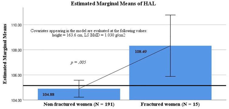
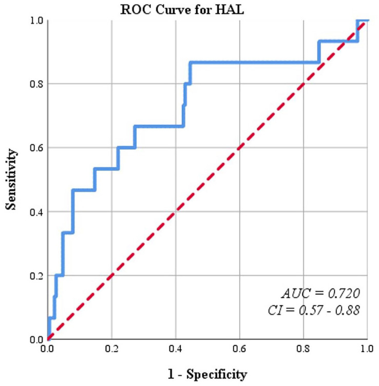
Results: The 15 fractured women displayed statistically greater HAL values compared to the 191 non-fractured subjects (109.43 ± 6.44 vs 104.81 ± 5.32 mm, p = 0.002), even though there were no significant differences in age, body mass index, or BMD. The difference in HAL remained significant after adjusting for lumbar spine (LS) BMD and height (108.49 ± 1.23 vs 104.88 ± 0.34 mm, p = 0.005). HAL proved to be a fair indicator of non-hip, non-vertebral cortical fractures (area under curve = 0.720, p = 0.003), with a sensitivity of 86.7% and a specificity of 55.5%.
Conclusion: HAL was positively associated with non-hip, non-vertebral cortical bone fragility fractures in young postmenopausal, clinically healthy women and had significantly greater values in the fractured subgroup even after adjusting for LS BMD and height.
虽然用双x线骨密度仪(DXA)测量骨密度(BMD)代表了骨质疏松症的诊断和治疗标准,但在骨密度不低的年轻患者中发生了大量脆性骨折。最近,临床风险工具包括髋轴长度(HAL),这是一个来自髋关节DXA扫描的几何参数,作为老年绝经后妇女髋部骨折的预测因子。本研究旨在评估年轻绝经后临床健康女性HAL与其他皮质骨折的关系。材料和方法:本研究回顾性分析了206例40-60岁的正常或超重高加索女性的月相DXA扫描,这些女性绝经时间小于10年,无继发性骨质疏松症,既往无骨质疏松症诊断或药物治疗,无髋部或椎体骨折史。结果:15名女性骨折患者的HAL值比191名非骨折患者高(109.43±6.44 vs 104.81±5.32 mm, p = 0.002),尽管年龄、体重指数和骨密度没有显著差异。调整腰椎骨密度(LS)和高度后,HAL的差异仍然显著(108.49±1.23 vs 104.88±0.34 mm, p = 0.005)。HAL被证明是非髋部、非椎体皮质骨折的合理指标(曲线下面积= 0.720,p = 0.003),敏感性为86.7%,特异性为55.5%。结论:HAL与绝经后临床健康的年轻女性的非髋部、非椎体皮质骨脆性骨折呈正相关,即使在调整了LS骨密度和身高后,HAL在骨折亚组中的价值也显著增加。
期刊介绍:
Therapeutic Advances in Endocrinology and Metabolism delivers the highest quality peer-reviewed articles, reviews, and scholarly comment on pioneering efforts and innovative studies across all areas of endocrinology and metabolism.

 求助内容:
求助内容: 应助结果提醒方式:


