Jowan Al-Nusair, Thomas Wright, Adamsegd Gebremedhen, Howide Eldib, Mohamed Alshal, Maria Tirona
{"title":"The Imitation Game: Melanoma Metastasis Poses as a Primary Breast Tumor.","authors":"Jowan Al-Nusair, Thomas Wright, Adamsegd Gebremedhen, Howide Eldib, Mohamed Alshal, Maria Tirona","doi":"10.1177/23247096251345394","DOIUrl":null,"url":null,"abstract":"<p><p>Metastatic melanoma to the breast is a rare phenomenon often mistaken for primary breast cancer due to overlapping clinical and imaging characteristics. We report the case of a 51-year-old woman with a history of melanoma resected 7 years earlier, presenting with severe left hip pain and a 6-month history of a right breast lump. Imaging revealed extensive metastatic disease, including lesions in the femoral head, breast, lung, adrenal gland, and thoracic spine. Pathologic examination of the hip lesion obtained during total arthroplasty, as well as biopsies of the breast and lung, confirmed metastatic melanoma. Histology revealed pleomorphic tumor cells with necrosis, while immunohistochemical analysis demonstrated SOX10 and S100 positivity, confirming the diagnosis. Genetic testing identified microsatellite stability with a tumor mutational burden of 16 mutations per mega base. This case shows the importance of thorough cancer histories and the use of immunohistochemical staining to distinguish metastatic melanoma from primary breast malignancies. Despite timely diagnosis and intervention, the patient's condition deteriorated rapidly, reflecting the aggressive nature of metastatic melanoma. This case highlights the need for vigilance in patients with a history of melanoma presenting with new breast masses to ensure accurate diagnosis and appropriate management.</p>","PeriodicalId":16198,"journal":{"name":"Journal of investigative medicine high impact case reports","volume":"13 ","pages":"23247096251345394"},"PeriodicalIF":0.8000,"publicationDate":"2025-01-01","publicationTypes":"Journal Article","fieldsOfStudy":null,"isOpenAccess":false,"openAccessPdf":"https://www.ncbi.nlm.nih.gov/pmc/articles/PMC12126677/pdf/","citationCount":"0","resultStr":null,"platform":"Semanticscholar","paperid":null,"PeriodicalName":"Journal of investigative medicine high impact case reports","FirstCategoryId":"1085","ListUrlMain":"https://doi.org/10.1177/23247096251345394","RegionNum":0,"RegionCategory":null,"ArticlePicture":[],"TitleCN":null,"AbstractTextCN":null,"PMCID":null,"EPubDate":"2025/5/31 0:00:00","PubModel":"Epub","JCR":"Q3","JCRName":"MEDICINE, GENERAL & INTERNAL","Score":null,"Total":0}
引用次数: 0
Abstract
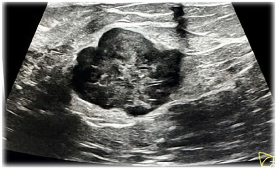
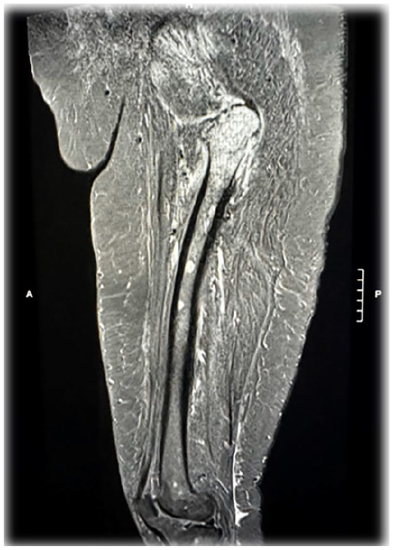
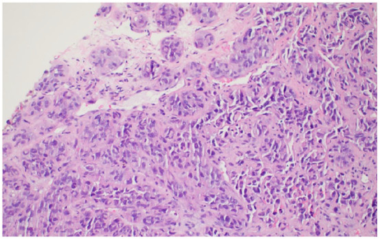
Metastatic melanoma to the breast is a rare phenomenon often mistaken for primary breast cancer due to overlapping clinical and imaging characteristics. We report the case of a 51-year-old woman with a history of melanoma resected 7 years earlier, presenting with severe left hip pain and a 6-month history of a right breast lump. Imaging revealed extensive metastatic disease, including lesions in the femoral head, breast, lung, adrenal gland, and thoracic spine. Pathologic examination of the hip lesion obtained during total arthroplasty, as well as biopsies of the breast and lung, confirmed metastatic melanoma. Histology revealed pleomorphic tumor cells with necrosis, while immunohistochemical analysis demonstrated SOX10 and S100 positivity, confirming the diagnosis. Genetic testing identified microsatellite stability with a tumor mutational burden of 16 mutations per mega base. This case shows the importance of thorough cancer histories and the use of immunohistochemical staining to distinguish metastatic melanoma from primary breast malignancies. Despite timely diagnosis and intervention, the patient's condition deteriorated rapidly, reflecting the aggressive nature of metastatic melanoma. This case highlights the need for vigilance in patients with a history of melanoma presenting with new breast masses to ensure accurate diagnosis and appropriate management.
期刊介绍:
The AFMR is committed to enhancing the training and career development of our members and to furthering its mission to facilitate the conduct of research to improve medical care. Case reports represent an important avenue for trainees (interns, residents, and fellows) and early-stage faculty to demonstrate productive, scholarly activity.

 求助内容:
求助内容: 应助结果提醒方式:


