Sustained Enlargement in Vagus and Sural Nerve Cross-Sectional Areas in Fibromyalgia: A Longitudinal Study
Abstract
Background and Purpose
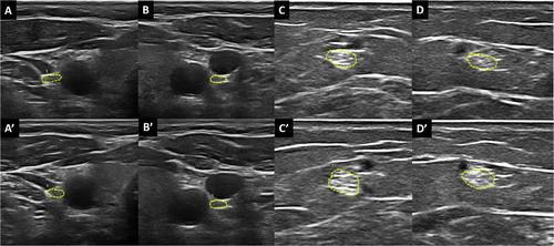
Fibromyalgia (FM) is a complex condition with unclear pathophysiology. While central sensitization is commonly accepted as the predominant cause of pain symptoms, numerous evidences suggest a role for the peripheral nervous system, particularly small fiber neuropathy. Previous studies have documented that patients with FM show an increased cross-sectional area (CSA) of some nerves, including the vagus and sural nerves, detectable via ultrasound (US). The purpose of this study is to assess whether the CSA increase persists over time and to investigate potential correlations between nerve dimensions and clinical variables.
Methods
This study involved 32 female patients with FM and 20 healthy controls, both evaluated at baseline and after 24 months. Participants completed clinimetric questionnaires addressing disease severity, neuropathic pain features, and autonomic dysfunction, while US measurements of the vagus and sural nerves' CSA were taken. Differences in CSA variation were assessed with student's t-test and chi-square, and the Pearson's correlation coefficient tested relationships between nerve dimensions and clinimetric scores.
Results
CSA values were higher in FM patients compared to controls at both baseline and after 24 months, although no significant differences in CSA changes were found over time. Pearson's correlation revealed some associations between nerve dimensions and clinimetric scores, suggesting potential relationships that require further investigation.
Conclusions
FM patients exhibit persistent increases in the vagus and sural nerves CSAs. Further studies are needed to better understand the clinical significance of these findings and the role of US assessment as a tool for detecting nerve alterations in FM.

 求助内容:
求助内容: 应助结果提醒方式:


