Pedunculated focal nodular hyperplasia: a case report, case series, and in-depth surgical, radiological, and histological analysis of a rare phenomenon.
Taylor Strange, Joseph M Gosnell, Peeyush Bhargava, Abdulrahman Al Harbi, Luca Cicalese, Heather L Stevenson
{"title":"Pedunculated focal nodular hyperplasia: a case report, case series, and in-depth surgical, radiological, and histological analysis of a rare phenomenon.","authors":"Taylor Strange, Joseph M Gosnell, Peeyush Bhargava, Abdulrahman Al Harbi, Luca Cicalese, Heather L Stevenson","doi":"10.1186/s13000-025-01661-y","DOIUrl":null,"url":null,"abstract":"<p><strong>Background: </strong>Focal nodular hyperplasia (FNH) is a benign hepatic lesion that rarely presents as an exophytic mass attached by a fibrous stalk (termed pedunculated FNH). This variation poses a challenge to clinicians, with atypical symptoms and imaging.</p><p><strong>Case presentation: </strong>We describe a 33-year-old female who underwent excision of a pedunculated FNH. On gross examination, the lesion was lobular and vascular with homogenous tan-brown surfaces. Histological examination showed loss of normal liver architecture, abnormal intervening fibrous tracts, dysplastic arteries, and focal steatosis. Immunohistochemical staining with glutamine synthetase resulted in a branching, or \"map-like\" pattern. These findings were consistent with focal nodular hyperplasia. One of the most sensitive imaging techniques for diagnosing this lesion involves magnetic resonance imaging (MRI) with contrast, which discloses a homogenous mass that is hyperintense during the arterial phase with gradual decrease in intensity during the venous and equilibrium phases. The central stellate scar will often remain hyperintense for a prolonged period of time. On histology, normal hepatic architecture is lost to abnormal fibrotic bands and a characteristic stellate scar. Immunohistochemistry with glutamine synthetase uniquely highlights a map-like pattern that is not seen in other liver lesions.</p><p><strong>Conclusions: </strong>Due to its atypical presentation and increased risk of complications compared to its intrahepatic counterpart, pedunculated FNH brings unique challenges for diagnosis and therapy. Proper identification of pedunculated FNH is critical for appropriate treatment. Our case highlights the importance of radiological and histopathological studies to accurately identify this lesion, as well as the benefits of surgical removal to prevent serious complications.</p>","PeriodicalId":11237,"journal":{"name":"Diagnostic Pathology","volume":"20 1","pages":"69"},"PeriodicalIF":2.3000,"publicationDate":"2025-05-30","publicationTypes":"Journal Article","fieldsOfStudy":null,"isOpenAccess":false,"openAccessPdf":"https://www.ncbi.nlm.nih.gov/pmc/articles/PMC12125829/pdf/","citationCount":"0","resultStr":null,"platform":"Semanticscholar","paperid":null,"PeriodicalName":"Diagnostic Pathology","FirstCategoryId":"3","ListUrlMain":"https://doi.org/10.1186/s13000-025-01661-y","RegionNum":3,"RegionCategory":"医学","ArticlePicture":[],"TitleCN":null,"AbstractTextCN":null,"PMCID":null,"EPubDate":"","PubModel":"","JCR":"Q2","JCRName":"PATHOLOGY","Score":null,"Total":0}
引用次数: 0
Abstract
Background: Focal nodular hyperplasia (FNH) is a benign hepatic lesion that rarely presents as an exophytic mass attached by a fibrous stalk (termed pedunculated FNH). This variation poses a challenge to clinicians, with atypical symptoms and imaging.
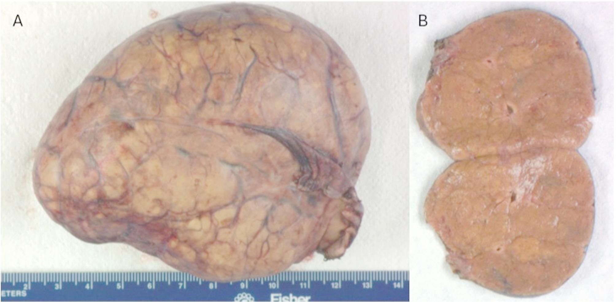
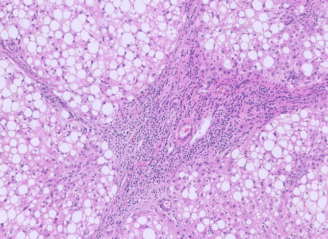
Case presentation: We describe a 33-year-old female who underwent excision of a pedunculated FNH. On gross examination, the lesion was lobular and vascular with homogenous tan-brown surfaces. Histological examination showed loss of normal liver architecture, abnormal intervening fibrous tracts, dysplastic arteries, and focal steatosis. Immunohistochemical staining with glutamine synthetase resulted in a branching, or "map-like" pattern. These findings were consistent with focal nodular hyperplasia. One of the most sensitive imaging techniques for diagnosing this lesion involves magnetic resonance imaging (MRI) with contrast, which discloses a homogenous mass that is hyperintense during the arterial phase with gradual decrease in intensity during the venous and equilibrium phases. The central stellate scar will often remain hyperintense for a prolonged period of time. On histology, normal hepatic architecture is lost to abnormal fibrotic bands and a characteristic stellate scar. Immunohistochemistry with glutamine synthetase uniquely highlights a map-like pattern that is not seen in other liver lesions.
Conclusions: Due to its atypical presentation and increased risk of complications compared to its intrahepatic counterpart, pedunculated FNH brings unique challenges for diagnosis and therapy. Proper identification of pedunculated FNH is critical for appropriate treatment. Our case highlights the importance of radiological and histopathological studies to accurately identify this lesion, as well as the benefits of surgical removal to prevent serious complications.
期刊介绍:
Diagnostic Pathology is an open access, peer-reviewed, online journal that considers research in surgical and clinical pathology, immunology, and biology, with a special focus on cutting-edge approaches in diagnostic pathology and tissue-based therapy. The journal covers all aspects of surgical pathology, including classic diagnostic pathology, prognosis-related diagnosis (tumor stages, prognosis markers, such as MIB-percentage, hormone receptors, etc.), and therapy-related findings. The journal also focuses on the technological aspects of pathology, including molecular biology techniques, morphometry aspects (stereology, DNA analysis, syntactic structure analysis), communication aspects (telecommunication, virtual microscopy, virtual pathology institutions, etc.), and electronic education and quality assurance (for example interactive publication, on-line references with automated updating, etc.).


 求助内容:
求助内容: 应助结果提醒方式:


