Amanda Beatriz Rodriguez, Berceste Guler Ayyildiz, Halil Ayyildiz, Aniruddh Narvekar, Mohammed H Elnagar, Michael Schmerman, Grace Viana, Salvador Nares, Tolga Fikret Tözüm
{"title":"CBCT Validation Study on Intraclass Correlation for Linear Measurements in Peri-implantitis: an Observational Study.","authors":"Amanda Beatriz Rodriguez, Berceste Guler Ayyildiz, Halil Ayyildiz, Aniruddh Narvekar, Mohammed H Elnagar, Michael Schmerman, Grace Viana, Salvador Nares, Tolga Fikret Tözüm","doi":"10.5037/jomr.2025.16104","DOIUrl":null,"url":null,"abstract":"<p><strong>Objectives: </strong>This retrospective observational study aimed to evaluate the reliability and reproducibility of intra-examiner and inter-examiner bone loss measurements in cone-beam computed tomography images over time, on patients with peri-implant defects using two cone-beam computed tomography software programs: 3D Slicer and Dolphin Imaging.</p><p><strong>Material and methods: </strong>Baseline images were oriented based on implant location and aligned with the palatal or Go-Me plane. Two CBCT volumes were imported and superimposed using landmark-based and surface-based methods, with accuracy assessed through 3D and 2D matching. Measurements of implant diameter, length, and bone thickness at 0, 1, 3, and 5 mm intervals were taken at two-time points by three independent examiners, with reliability assessed using intra-class correlation coefficients.</p><p><strong>Results: </strong>Twenty measurements per 14 cases were evaluated. Each examiner conducted 1,120 measurements with a cumulative total of 3,360 measurements assessed. Significant differences in measurement times were observed, with 3D Slicer requiring more time for superimposition tasks (P < 0.001). Both software programs, however, demonstrated high reliability (intraclass correlation coefficient > 0.80) in inter- and intra-examiner agreement across various bone measurements.</p><p><strong>Conclusions: </strong>Findings emphasize that the high reliability observed with the software and superimposition techniques is directly linked to the calibration and training exercises conducted with the examiners before the study. Dolphin's Imaging automated superimposition was significantly faster than 3D Slicer's manual approach, 3D Slicer offered superior image quality and better differentiation of bone outlines. Both software demonstrated effectiveness in delivering consistent and reproducible measurements, with significant implications for clinical and research applications in implant dentistry.</p>","PeriodicalId":53254,"journal":{"name":"eJournal of Oral Maxillofacial Research","volume":"16 1","pages":"e4"},"PeriodicalIF":1.0000,"publicationDate":"2025-03-31","publicationTypes":"Journal Article","fieldsOfStudy":null,"isOpenAccess":false,"openAccessPdf":"https://www.ncbi.nlm.nih.gov/pmc/articles/PMC12118470/pdf/","citationCount":"0","resultStr":null,"platform":"Semanticscholar","paperid":null,"PeriodicalName":"eJournal of Oral Maxillofacial Research","FirstCategoryId":"1085","ListUrlMain":"https://doi.org/10.5037/jomr.2025.16104","RegionNum":0,"RegionCategory":null,"ArticlePicture":[],"TitleCN":null,"AbstractTextCN":null,"PMCID":null,"EPubDate":"2025/1/1 0:00:00","PubModel":"eCollection","JCR":"Q3","JCRName":"DENTISTRY, ORAL SURGERY & MEDICINE","Score":null,"Total":0}
引用次数: 0
Abstract
Objectives: This retrospective observational study aimed to evaluate the reliability and reproducibility of intra-examiner and inter-examiner bone loss measurements in cone-beam computed tomography images over time, on patients with peri-implant defects using two cone-beam computed tomography software programs: 3D Slicer and Dolphin Imaging.
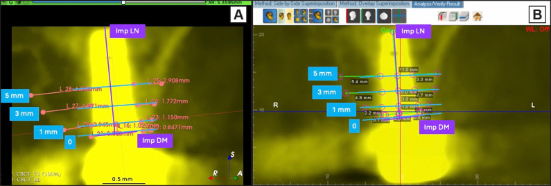
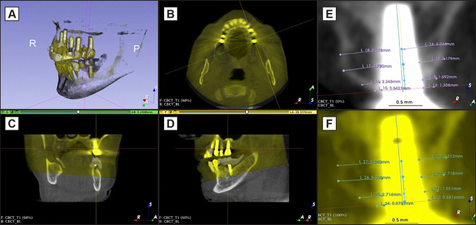
Material and methods: Baseline images were oriented based on implant location and aligned with the palatal or Go-Me plane. Two CBCT volumes were imported and superimposed using landmark-based and surface-based methods, with accuracy assessed through 3D and 2D matching. Measurements of implant diameter, length, and bone thickness at 0, 1, 3, and 5 mm intervals were taken at two-time points by three independent examiners, with reliability assessed using intra-class correlation coefficients.
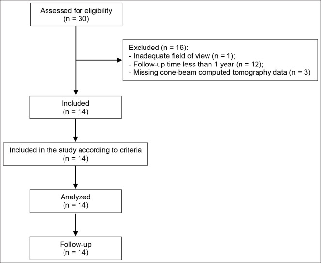
Results: Twenty measurements per 14 cases were evaluated. Each examiner conducted 1,120 measurements with a cumulative total of 3,360 measurements assessed. Significant differences in measurement times were observed, with 3D Slicer requiring more time for superimposition tasks (P < 0.001). Both software programs, however, demonstrated high reliability (intraclass correlation coefficient > 0.80) in inter- and intra-examiner agreement across various bone measurements.
Conclusions: Findings emphasize that the high reliability observed with the software and superimposition techniques is directly linked to the calibration and training exercises conducted with the examiners before the study. Dolphin's Imaging automated superimposition was significantly faster than 3D Slicer's manual approach, 3D Slicer offered superior image quality and better differentiation of bone outlines. Both software demonstrated effectiveness in delivering consistent and reproducible measurements, with significant implications for clinical and research applications in implant dentistry.

 求助内容:
求助内容: 应助结果提醒方式:


