Barbara Lenz, Martina Stirn, Elke-Astrid Atzpodien, Annamaria Braendli-Baiocco, Anna Maria Giusti, Kerstin Hahn, Vanessa Schumacher
{"title":"Ring hemorrhages in the central nervous system of severely anemic cynomolgus monkeys.","authors":"Barbara Lenz, Martina Stirn, Elke-Astrid Atzpodien, Annamaria Braendli-Baiocco, Anna Maria Giusti, Kerstin Hahn, Vanessa Schumacher","doi":"10.1177/03009858251343012","DOIUrl":null,"url":null,"abstract":"<p><p>Ring hemorrhages are characteristic ring-shaped lesions in the parenchyma of the central nervous system (CNS) surrounding small blood vessels and are typically reported to occur in white matter of the brain. In humans, they are seen in various diseases, including pernicious anemia, cerebral fat embolism, and cerebral malaria. Ring hemorrhages are also sporadically seen in the CNS of animals and have been previously reported in 4 nonhuman primate (NHP) species with various forms of anemia. Here we present the results of 4 preclinical toxicity studies testing 4 drug candidates of different modalities, targets, mechanisms of action, and indications in NHPs (cynomolgus monkeys). Within each study, ring hemorrhages were observed associated with severe anemia in affected animals and were in line with those previously reported. They occurred in gray matter and occasionally white matter of the brain and spinal cord; in the brain, the thalamus, basal ganglia, and cerebellum were particularly affected. Lesions comprised a central eosinophilic core, sometimes with small blood vessels or eosinophilic to slightly basophilic amorphous material in the center, surrounded by red blood cells and/or microglial cells or microglia aggregates. Fibrin staining confirmed the presence of fibrin in the central core. No commonality in type/cause of anemia was noted; in one study, the anemia was considered a spontaneous (non-treatment-related) finding. A thorough examination of the brain is therefore recommended in the presence of anemic conditions in animals. Pathologists should be aware of this finding and its relationship with anemia when assessing associations with diseases or drug candidates.</p>","PeriodicalId":23513,"journal":{"name":"Veterinary Pathology","volume":" ","pages":"3009858251343012"},"PeriodicalIF":1.7000,"publicationDate":"2025-05-30","publicationTypes":"Journal Article","fieldsOfStudy":null,"isOpenAccess":false,"openAccessPdf":"https://www.ncbi.nlm.nih.gov/pmc/articles/PMC12185903/pdf/","citationCount":"0","resultStr":null,"platform":"Semanticscholar","paperid":null,"PeriodicalName":"Veterinary Pathology","FirstCategoryId":"97","ListUrlMain":"https://doi.org/10.1177/03009858251343012","RegionNum":2,"RegionCategory":"农林科学","ArticlePicture":[],"TitleCN":null,"AbstractTextCN":null,"PMCID":null,"EPubDate":"","PubModel":"","JCR":"Q2","JCRName":"PATHOLOGY","Score":null,"Total":0}
引用次数: 0
Abstract
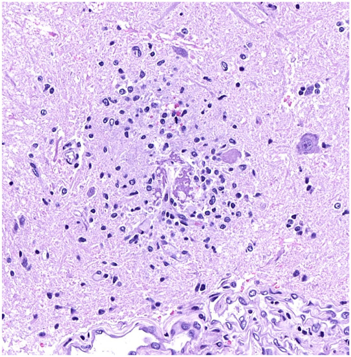
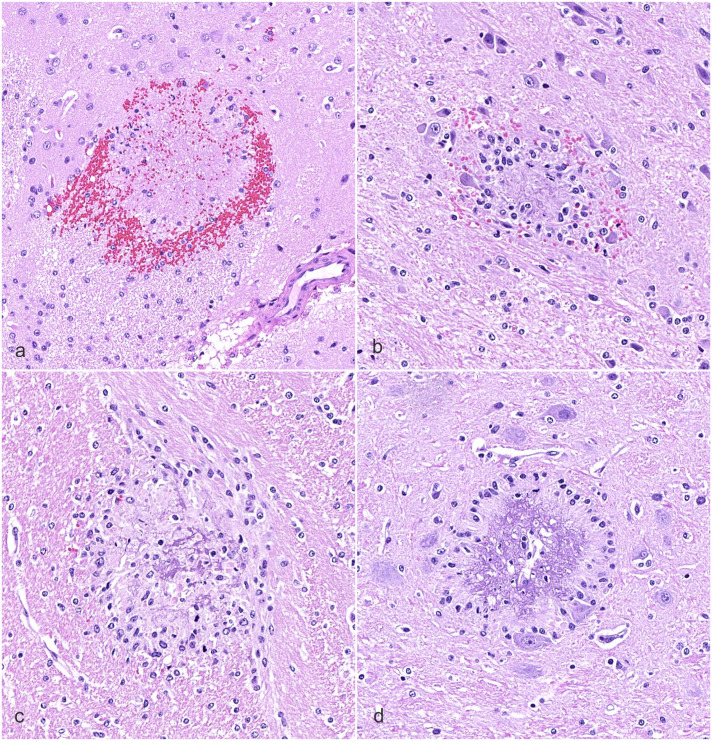
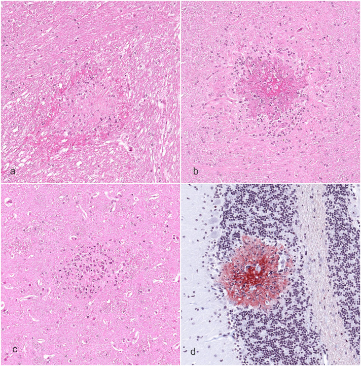
Ring hemorrhages are characteristic ring-shaped lesions in the parenchyma of the central nervous system (CNS) surrounding small blood vessels and are typically reported to occur in white matter of the brain. In humans, they are seen in various diseases, including pernicious anemia, cerebral fat embolism, and cerebral malaria. Ring hemorrhages are also sporadically seen in the CNS of animals and have been previously reported in 4 nonhuman primate (NHP) species with various forms of anemia. Here we present the results of 4 preclinical toxicity studies testing 4 drug candidates of different modalities, targets, mechanisms of action, and indications in NHPs (cynomolgus monkeys). Within each study, ring hemorrhages were observed associated with severe anemia in affected animals and were in line with those previously reported. They occurred in gray matter and occasionally white matter of the brain and spinal cord; in the brain, the thalamus, basal ganglia, and cerebellum were particularly affected. Lesions comprised a central eosinophilic core, sometimes with small blood vessels or eosinophilic to slightly basophilic amorphous material in the center, surrounded by red blood cells and/or microglial cells or microglia aggregates. Fibrin staining confirmed the presence of fibrin in the central core. No commonality in type/cause of anemia was noted; in one study, the anemia was considered a spontaneous (non-treatment-related) finding. A thorough examination of the brain is therefore recommended in the presence of anemic conditions in animals. Pathologists should be aware of this finding and its relationship with anemia when assessing associations with diseases or drug candidates.
期刊介绍:
Veterinary Pathology (VET) is the premier international publication of basic and applied research involving domestic, laboratory, wildlife, marine and zoo animals, and poultry. Bridging the divide between natural and experimental diseases, the journal details the diagnostic investigations of diseases of animals; reports experimental studies on mechanisms of specific processes; provides unique insights into animal models of human disease; and presents studies on environmental and pharmaceutical hazards.
 求助内容:
求助内容: 应助结果提醒方式:


